
Illustrated Encyclopedia of Human Anatomic Variation: Opus II: Cardiovascular System
Ronald A. Bergman, PhD
Adel K. Afifi, MD, MS
Ryosuke Miyauchi, MD
Peer Review Status: Internally Peer Reviewed
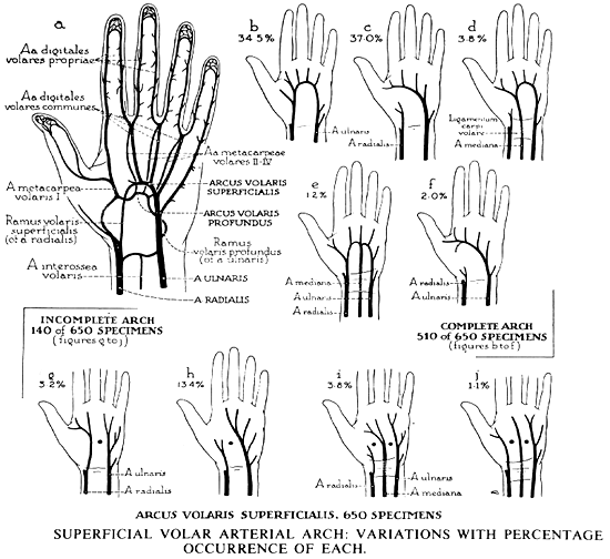
Types of superficial volar (palmer) arterial arch (b-j) encountered in 650 dissections of the hand, together with the "textbook normal", (a). Shown schematically, with percentage occurrence of each of the nine types. Variations (b-f), encountered in 510 specimens, in which the arch was complete; patterns (g-j), found in 140 hands, in which the arch was incomplete - in the areas indicated by a marker or markers.
a= So-called "textbook normal"
b= Schematized form of the complete radioulnar communication that was present in more than one-third of the total number of extremities studied.
c= Transpalmar arciform continuation of the ulnar artery with a full complement of common volar digital branches. This type occurs more frequently, 37% of cases, than the "textbook normal or standard" form.
d= Arciform arrangement to which the contributors are ulnar and median arteries - the median replacing the radial of the type shown in a and b. The ulnomedian type initiates the series of patterns whose frequencies of occurrence show a steep decline from the most common varieties; namely those in b and e.
e= An arch, to which all three arteries contribute. Here a median communication is sent to the center of the arch formed by anastomoses of the radial and ulnar arteries.
f= A transpalmar continuation of the ulnar artery (compare with c) receives a midpalmar contribution from the deep palmer arterial arch, not from the radial artery itself. In the remaining specimens, 140 out of a total of 650 (21.5%), the superficial arterial arch is incomplete, the area of interruption indicated in each diagram by a black dot.
g= In specimens of this type, the proper volar arteries are derived equally form the radial and ulnar arteries, without communication across the middle line of the hand.
h= The ulnar artery is the chief contributor to the set of digital vessels, supplying three and one-half digits; that is, toward the thumb, to include the ulnar aspect of the index finger.
i= In specimens of this type, the median artery reaches the hand to furnish digital arteries (compare with d and e) but without anastomosing the radial and ulnar arteries. Inclining toward the radial side of the hand, the median artery gives off a branch to the thumb.
j= In this variety of palmer arterial supply, the deviation of the three source-vessels toward the radial side of the hand is of lesser degree than in the preceding type; the branches of the common digital artery derived from the median artery pass only to the facing aspects of the index and middle fingers, the radial and ulnar arteries caring for the areas marginal thereto.
from Coleman and Anson, 1961.
Section Top | Title Page
Please send us comments by filling out our Comment Form.
All contents copyright © 1995-2024 the Author(s) and Michael P. D'Alessandro, M.D. All rights reserved.
"Anatomy Atlases", the Anatomy Atlases logo, and "A digital library of anatomy information" are all Trademarks of Michael P. D'Alessandro, M.D.
Anatomy Atlases is funded in whole by Michael P. D'Alessandro, M.D. Advertising is not accepted.
Your personal information remains confidential and is not sold, leased, or given to any third party be they reliable or not.
The information contained in Anatomy Atlases is not a substitute for the medical care and advice of your physician. There may be variations in treatment that your physician may recommend based on individual facts and circumstances.
URL: http://www.anatomyatlases.org/