
Illustrated Encyclopedia of Human Anatomic Variation: Opus II: Cardiovascular System
Ronald A. Bergman, PhD
Adel K. Afifi, MD, MS
Ryosuke Miyauchi, MD
Peer Review Status: Internally Peer Reviewed
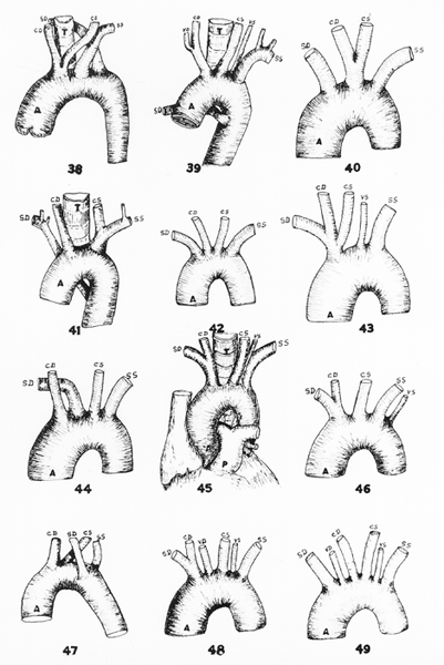
Fig. 38:This figure was drawn from a specimen of low origin of the right subclavian with both carotids springing from a common stem, which is in the Warren Museum (Harvard University).
Fig. 39:This figure, taken from Krause (1876), Fig. 115 (Mccartney, and Tiedemann, (1846), Fig. 6, Plate XXXIX, represents a case of low origin of the right subclavian artery with the right vertebral springing from the right common carotid abd left vertebral from the aortic arch.
Fig. 40:This figure, representing a case of common trunk for the carotids, was taken from Tiedemann (1822), Fig. 2, Plate III; copied by Quain (1844) as Fig. 4, Plate VII.
Fig. 41:This figure, representing a case of low right subclavian artery, with a common stem for the left carotid and subclavian, was taken from Tiedemann (1822), Fig. 6, Plate II: copied by Quain (1844) as Fig. 8, Plate VII. (Note also that the right subclavian artery will be either retrotracheal or retroesophageal.)
Fig. 42:This figure, representing a case in which all four branches spring separately from the arch, was taken from Tiedemann (1822), Fig. 3, Plate III; copied by Quain (1814) as Fig. 10, Plate VI.
Fig. 43:This figure, representing a case of the left common carotid arising from the innominate trunk and the left vertebral from the arch, was taken from Tiedemann (1822), Fig. 7, Plate II.
Fig. 44:This figure, representing a case of the right subclavian arising as the second branch from the arch, was taken from Tiedemann (1822), Fig. 4, Plate III; copied from Huber (1777), vol. 8, p. 75 and Fig. 3.
Fig. 45:This figure is adapted from a case of left vertebral arising from the aortic arch, which was found in our (Poynter's) dissecting rooms.
Fig. 46:This figure, representing a case in which the left vertebral is the last branch from the arch, was taken from Tiedemann (1822), Fig.10, Plate III; copied by Quain as Fig. 10, Plate VII.
Fig. 47:This figure, representing the right subclavian as the third branch from the arch, was reported by Walter (1785), p. 62, Fig. 5, Plate III; it was copied by both Quain and Tiedemann.
Fig. 48:This figure, representing five branches from the arch, was reported by Penada (1801), p. 44, and illustrated by Tiedemann (1822) as fig. 4, Plate IV.
Fig. 49:This figure, representing six branches from the arch, was taken from Tiedemann (1822), fig. 5, Plate IV; copied by Quain (1844) as fig. 15, Plate VII.
Abbreviations used in the figures:
A, aorta; AD, aorta descendens (descending aorta); C, carotis
(carotid); CD, a. carotis communis dextra (right common carotid); CS
a carotis sinistra (left common carotid); D, ductus arteriosus
(Botalli); EC, a. carotis externis (external carotid artery); IC, a
carotis internis (internal carotid artery); O, esophagus; P, a.
pulmonalis (pulmonary artery); SD, a subclavia dextra (right
subclavian artery); SS, a. subclavia sinistra (left subclavian
artery); T, trachea; TD, ductus thoracicus (thoracic duct); VD, a.
vertebralis dextra (right vertebral artery); VS, a vertebralis
sinistra (left vertebral artery).
from Poynter, 1916.
Section Top | Title Page
Please send us comments by filling out our Comment Form.
All contents copyright © 1995-2024 the Author(s) and Michael P. D'Alessandro, M.D. All rights reserved.
"Anatomy Atlases", the Anatomy Atlases logo, and "A digital library of anatomy information" are all Trademarks of Michael P. D'Alessandro, M.D.
Anatomy Atlases is funded in whole by Michael P. D'Alessandro, M.D. Advertising is not accepted.
Your personal information remains confidential and is not sold, leased, or given to any third party be they reliable or not.
The information contained in Anatomy Atlases is not a substitute for the medical care and advice of your physician. There may be variations in treatment that your physician may recommend based on individual facts and circumstances.
URL: http://www.anatomyatlases.org/