
Illustrated Encyclopedia of Human Anatomic Variation: Opus II: Cardiovascular System
Ronald A. Bergman, PhD
Adel K. Afifi, MD, MS
Ryosuke Miyauchi, MD
Peer Review Status: Internally Peer Reviewed
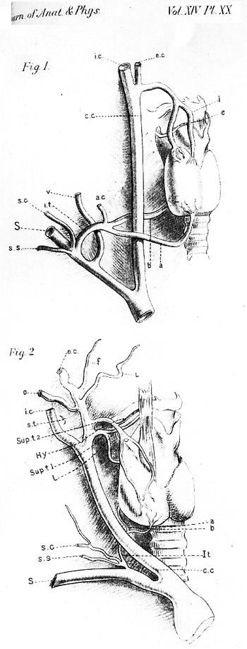
A.:"The right superior thyroid arose from the external carotid near its origin, and divided into medial and lateral branches. The inferior thyroid arose from the subclavian, gave rise to the ascending cervical, and immediately divided into superior and inferior branches. The former passed upwards and deep to the carotid sheath and beneath the right lobe of the thyroid gland, and distributed to the posterior, lower, and lateral part of that lobe. The latter passed in front of the carotid sheath at a more inferior level than the superior branch, and after a more prolonged course supplied the medial and inferior part of the right lobe of the gland, and the anterior surface."
B.: "Two right superior thyroid arteries arose from the medial part of the external carotid. The upper superior thyroid, which was larger, passed downward and medially, and divided into two branches near the superior border of the thyroid cartilage; of these, the medial branch, passed beneath the sternohyoid and over the thyrohyoid, and then downward over the thyroid gland, and distributed to the medial part of the anterior surface and the isthmus, and gave rise to a cricothyroid branch at the lower border of the thyroid cartilage. The lateral branch passed beneath the sternothyroid, and crossed the right lobe obliquely from above downward and inward, and distributed to the anterior surface, lateral to the medial branch, reaching as far as the lower third of the gland."
Abbreviations for both illustrations: a.c= ascending cervical artery; c.c= common carotid artery; e= superior thyroid artery, lateral branch; e.c= external carotid; f= facial artery; Hy= hyoid artery; I= superior thyroid artery, medial branch; ic= internal carotid artery; i.t (I.t)= inferior thyroid artery: a= anterior branch, and b= deep branch; L= lingual branch; o= occipital artery; S= subclavian artery, right; s.c= superficial cervical artery; s.s= suprascapular artery; Sup.t 1= superior thyroid artery, first; Sup.t 2= superior thyroid, second; V= vertebral artery.
Authors' note: Quain found three cases of double superior thyroid arteries in 292 cases. Tiedemann and Gruber also reported doubled superior thyroid arteries.
from Anderson, 1879.
Section Top | Title Page
Please send us comments by filling out our Comment Form.
All contents copyright © 1995-2024 the Author(s) and Michael P. D'Alessandro, M.D. All rights reserved.
"Anatomy Atlases", the Anatomy Atlases logo, and "A digital library of anatomy information" are all Trademarks of Michael P. D'Alessandro, M.D.
Anatomy Atlases is funded in whole by Michael P. D'Alessandro, M.D. Advertising is not accepted.
Your personal information remains confidential and is not sold, leased, or given to any third party be they reliable or not.
The information contained in Anatomy Atlases is not a substitute for the medical care and advice of your physician. There may be variations in treatment that your physician may recommend based on individual facts and circumstances.
URL: http://www.anatomyatlases.org/