
Illustrated Encyclopedia of Human Anatomic Variation: Opus II: Cardiovascular System
Ronald A. Bergman, PhD
Adel K. Afifi, MD, MS
Ryosuke Miyauchi, MD
Peer Review Status: Internally Peer Reviewed

"Anatomic variations of the sinuses of the dura mater, however infrequent, may present puzzling diagnostic and operative problems in the presence of thrombophlebitis. Some of the variations are extremely rare. In this paper a hitherto unknown anomaly of the sigmoid sinus will be reported and a short description of most of the published observations which are important for the otologist will be given. In view of the existing differences in the nomenclature, in this (report) the horizontal portion of the transverse sinus will be called the lateral sinus and the vertical portion will be called the sigmoid sinus."
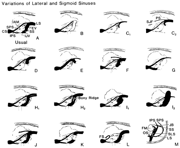
Abbreviations: CS, cavernous sinus; EV, mastoid emissary vein; FM, foramen magnum; FS, foramen spinosum; JAM, internal auditory meatus; UV, internal jugular vein; IPS, inferior petrosal sinus; JB, jugular bulb; LS, transverse sinus; OS, ophthalmopetrosal sinus; PS, petrosquamosal sinus; SJF, spurious jugular foramen; SLS, superior sagittal sinus; SPS, superior petrosal sinus; SS, sigmoid sinus.
A. Representation of the usual sinus region (intracranial view). Other drawings, from the same view, show anatomic variations of venous sinuses; B. lateral sinus absent; C1. small transverse sinus leaving endocranium through mastoid foremen; C2:. persistent petrosquamous sinus present; D. petrous bone infantile; E. superior petrosal sinus passing through mastoid foramen; F. sigmoid sinus ending in a blind pouch; G. complete absence of sigmoid sinus. Diagrammatic representations of the other (variations) of the region of the sinuses (intracranial view). H1. H2. duplications of lateral and sigmoid sinuses; I. hernia-like bulging of outer wall: (1) in upper part of region of knee and (2) with jugular bulb missing; J. persistent petrosquamous sinus draining lateral sinus; K. cortical layer thinned to paper-like sheet by enlarged sigmoid sinus; L. superior petrosal sinus turning downward into foramen spinosum of middle fossa; M. superior longitudinal sinus continuing directly with jugular bulb.
redrawn from Waltner, 1944
Section Top | Title Page
Please send us comments by filling out our Comment Form.
All contents copyright © 1995-2024 the Author(s) and Michael P. D'Alessandro, M.D. All rights reserved.
"Anatomy Atlases", the Anatomy Atlases logo, and "A digital library of anatomy information" are all Trademarks of Michael P. D'Alessandro, M.D.
Anatomy Atlases is funded in whole by Michael P. D'Alessandro, M.D. Advertising is not accepted.
Your personal information remains confidential and is not sold, leased, or given to any third party be they reliable or not.
The information contained in Anatomy Atlases is not a substitute for the medical care and advice of your physician. There may be variations in treatment that your physician may recommend based on individual facts and circumstances.
URL: http://www.anatomyatlases.org/