
Illustrated Encyclopedia of Human Anatomic Variation: Opus II: Cardiovascular System
Ronald A. Bergman, PhD
Adel K. Afifi, MD, MS
Ryosuke Miyauchi, MD
Peer Review Status: Internally Peer Reviewed
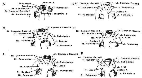
A: Right-sided aortic arch and upper portion of the descending
aorta, and left-sided ductus arteriosus in which the ductus
arteriosis passed behind the esophagus to insert into an aortic
diverticulum.
B: Right-sided aortic arch and upper portion of the descending aorta,
and left-sided ductus arteriosus in which the ductus arteriosus
inserted into a left subclavian artery which arose as the fourth
branch from the aortic arch.
C: Left-sided descending aorta and left-sided ductus arteriosus.
D: Left-sided descending aorta and right-sided ductus arteriosus.
E. Right-sided upper portion of the descending aorta and right-sided
ductus arteriosus.
F. Right-sided upper portion of the descending aorta and left-sided
ductus arteriosus.
Kirklin, Clagett, and Edwards have provided the following classificiation of anomalies of aortic arch and a detailed discussion of these anomalies is givien by Edwards, J.E. Anomalies of the derivatives of the aortic arch system. Med. Clin. North Am. 32: 925-949, 1948.
Classification of Anomalies of Aortic Arch
B. Double aortic arch with partial atresia of one arch.
C. Right-sided aortic arch with retroesophageal segment and
left-sided descending aorta.
D. Left-sided aortic arch.
E. Right-sided ductus arteriosus arising from right pulmonary artery. All possibilities occurring under A could occur here.
2. Double aortic arch with partial atresia of one arch.
3. Left-sided aortic arch with retroesophageal segment and
right-sided upper portion of the sescending aorta.
4. Right-sided aortic arch.
Redrawn from Kirklin, J.W. and O.T. Clagett. Vascular "rings" producing respiratory obstruction in infants. Proc. Staff Meeting Mayo Clin. 25:360-367, 1950.
Section Top | Title Page
Please send us comments by filling out our Comment Form.
All contents copyright © 1995-2024 the Author(s) and Michael P. D'Alessandro, M.D. All rights reserved.
"Anatomy Atlases", the Anatomy Atlases logo, and "A digital library of anatomy information" are all Trademarks of Michael P. D'Alessandro, M.D.
Anatomy Atlases is funded in whole by Michael P. D'Alessandro, M.D. Advertising is not accepted.
Your personal information remains confidential and is not sold, leased, or given to any third party be they reliable or not.
The information contained in Anatomy Atlases is not a substitute for the medical care and advice of your physician. There may be variations in treatment that your physician may recommend based on individual facts and circumstances.
URL: http://www.anatomyatlases.org/