
Illustrated Encyclopedia of Human Anatomic Variation: Opus II: Cardiovascular System
Ronald A. Bergman, PhD
Adel K. Afifi, MD, MS
Ryosuke Miyauchi, MD
Peer Review Status: Internally Peer Reviewed
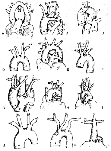
A: This figure, representing a case of true double aortic arch, was taken from Tiedemann (1822), Fig. 7, Plate IV, representing a case described by Malacarne (1788), Pt. 2, p. 119; it was copied by Quain (1844) as Fig. 8, Plate V, and by Krause (1876) as Fig. 108a.
B: This figure, representing a case in which the arch alone is double, was taken from Tiedemann (1822), Fig.6, Plate IV, representing a case described by Hommel (1737). It is referred to in Haller Elementary Physiology, Tom. 2, p. 162, and copied by Quain (1844) asFig.7, Plate V, and by Krause (1876) as Fig. 108b.
C: This figure, representing a case of right aortic arch with the left subclavian artery as the last branch and the ductus arteriosus patent, was taken from Quain (1844), Fig. 2, Plate VII.
D: This figure, representing a case of right aortic arch with a left innominate trunk, was taken from Tiedemann (1822), Fig. 9, Plate IV.
E: This figure, representing a case of persistence of both the right and left ductus arteriosus, was taken from Breschet (1826), Fig. 9, Plate 1.
F: This figure, representing the more or less common anomaly of left carotid artery springing from the innominate trunk, was copied from Tiedemann (1822), Fig. 5, Plate 11.
G: This figure, representing a case of right aortic arch, with the left subclavian artery as the last branch and the right vertebral springing from the arch, was taken from Abbott (1892), Fig-L H: This figure, representing a case of atresia of the pulmonary artery, was taken from Keith (1909), Fig.4; the pulmonary artery leaves the heart as a fibrous cord but rapidly enlarges to about normal, the ductus arteriosus is patent.
I: This figure, representing a case of thyroidea ima springing from the arch, was taken from Neubauer (1786), Fig. 2, Plate VII.
J: This figure, representing a case of bi-innominate trunks, was taken from Tiedemann (1822), Fig.4, Plate 11, and copied by Quain (1844), Fig. 9, Plate VI.
K: This figure was drawn from a specimen of left common carotid springing from the innominate, which is in the Warren Museum (Harvard University).
L: This figure, representing a single branch from the aortic arch, was taken from Tiedemann (1822), Fig. 3, Plate II, illustrating the case of Klinz (1793), p. 273. It was copied by Quain (1844), Fig. 6, Plate V.
Section Top | Title Page
Please send us comments by filling out our Comment Form.
All contents copyright © 1995-2024 the Author(s) and Michael P. D'Alessandro, M.D. All rights reserved.
"Anatomy Atlases", the Anatomy Atlases logo, and "A digital library of anatomy information" are all Trademarks of Michael P. D'Alessandro, M.D.
Anatomy Atlases is funded in whole by Michael P. D'Alessandro, M.D. Advertising is not accepted.
Your personal information remains confidential and is not sold, leased, or given to any third party be they reliable or not.
The information contained in Anatomy Atlases is not a substitute for the medical care and advice of your physician. There may be variations in treatment that your physician may recommend based on individual facts and circumstances.
URL: http://www.anatomyatlases.org/