
Illustrated Encyclopedia of Human Anatomic Variation: Opus II: Cardiovascular System
Ronald A. Bergman, PhD
Adel K. Afifi, MD, MS
Ryosuke Miyauchi, MD
Peer Review Status: Internally Peer Reviewed
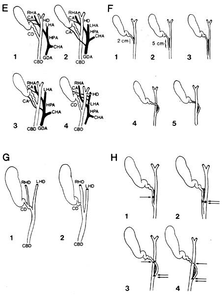
RHA, Right hepatic artery; LHA, left hepatic artery; HPA, main hepatic artery; CHA, common hepatic trunk; GDA, gastroduodenal artery; CA, cystic artery; HD, main hepatic duct; CD, cystic duct; CBD, common bile duct; SPDA, superior pancreatoduodenal artery.
E: Various modes of origin of double cystic arteries. 1: In 8% of the cases having two cystic arteries, both vessels arise from the right hepatic artery.
2: In 2% one artery arises from the right hepatic artery.
3: In 1% one artery arises from the right hepatic and the other from the main hepatic (hepatica propria) artery.
4: In 1% both vessels arise from the right hepatic artery.
F: Variations in the mode of union of the cystic and main hepatic ducts. 1: Normal (75%) unite at acute angle. Terminal 2 cm paralleled and held together firmly by fibrous tissue.
2: Short parallel type. Parallel for 5 cm or more as far as upper border of pancreas.
3: Long parallel type. Parallel almost throughout course, i.e., to within 1/2--l cm from ampulla of Vater.
2, 3: Together occurred in 17%.
4, 5: Anterior and posterior spiral types occurred in 8%. Note how cystic duct winds around anterior (or posterior) surface of the hepatic duct to enter its left border.
G: Anomalies in the right hepatic duct. 1: Right hepatic duct empties into cystic duct.
2: Cystic duct empties into the right hepatic duct.
H: Possible locations of calculi in cases with anomalies of the mode of union of the cystic and hepatic ducts. 1: Calculus in cystic duct of short parallel type. Can compress hepatic duct and cause same symptom as calculus in that duct.
2: Calculi in long parallel type of ducts. Could cause great technical difficulty in removal, with possible injury of ducts.
3: Calculi in spiral cystic duct. Very puzzling clinical picture if one (single arrow) compressed hepatic duct and other obstructed a cystic duct (double arrow) emptying into hepatic duct on its left side.
4: Similar possibilities from clinical and operative standpoint as in 3.
Redrawn from Eisendrath, D.N. The clinical importance of anatomical anomalies in biliary surgery. Boston Med. Surg. J. 182:573-578, 1920.
Section Top | Title Page
Please send us comments by filling out our Comment Form.
All contents copyright © 1995-2024 the Author(s) and Michael P. D'Alessandro, M.D. All rights reserved.
"Anatomy Atlases", the Anatomy Atlases logo, and "A digital library of anatomy information" are all Trademarks of Michael P. D'Alessandro, M.D.
Anatomy Atlases is funded in whole by Michael P. D'Alessandro, M.D. Advertising is not accepted.
Your personal information remains confidential and is not sold, leased, or given to any third party be they reliable or not.
The information contained in Anatomy Atlases is not a substitute for the medical care and advice of your physician. There may be variations in treatment that your physician may recommend based on individual facts and circumstances.
URL: http://www.anatomyatlases.org/