
Illustrated Encyclopedia of Human Anatomic Variation: Opus II: Cardiovascular System
Ronald A. Bergman, PhD
Adel K. Afifi, MD, MS
Ryosuke Miyauchi, MD
Peer Review Status: Internally Peer Reviewed
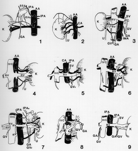
AA, Abdominal aorta; ARV, accessory renal vein; CA, celiac artery; CIA, common iliac artery; CIV, common iliac vein; GA, gonadal artery, GVv, gonadal veins; HV, hemiazygous vein; IMA, inferior mesenteric artery; IPA, inferior phrenic artery; IPV, inferior phrenic vein; ISA, inferior suprarenal artery; ISAa, inferior suprarenal arteries; IVC, inferior vena cava; K, kidney; MSV, middle sacral vein; RA, renal artery; SSA, superior suprarenal artery; SV, suprarenal vein; U, ureter; VL, venous Loops.
1: Right side, arteries: A supernumerary renal artery, coursing transversely as a constituent of the pedicle, gives off 3 suprarenal twigs and, in midcourse, an internal spermatic artery. The main artery sends a suprarenal branch cranialward, and accessory polar ramus lateralward; from the latter, in turn, a suprarenal twig arises. The right phrenic gives off three small suprarenal twigs.
2: Right, arteries: Three renal arteries; from the lowermost, in addition to a hilar branch, 1 branch enters the inferior extremity. Branches to the superior extremity arise from the intermediate one of the 3 renal arteries and from the inferior phrenic.
3: Right, arteries: Four renal arteries of approximately equal caliber. The superior 2 arise near the origin of the superior mesenteric, pass behind the vena cava; the next artery is precaval, enters the lower part of the hilus. The inferior member arises at the level of the inferior mesenteric, is also precaval, reaches the lower extremity just beyond the true hilus. Veins: The superior 1 of 4 renal veins is small, passes from the superior extremity; the next appears from behind the most cranial of the right renal arteries; the third passes along the caudal margin of the latter artery; the fourth vein, placed between the preaortic renal arteries, enters the vena cava by a short channel which it shares with the internal spermatic vein. Left, arteries: A single renal artery, concealed by the renal vein. Veins: A single renal vein, of preaortic course, crosses behind the superior mesenteric artery; it receives suprarenal, internal spermatic, and parietal vein.
4: Left side, arteries: Four renal arteries, 3 of them small. The first supplies the superior extremity; the second, the extremity and hilus; the third, the inferior aspect of the hilus; the fourth, the inferior extremity. Veins: A single large vessel.
5: Left, arteries: Two segmentally arranged renal arteries. Veins: A single large renal vein which receives a single suprarenal vein above, a parietal tributary behind (out of view), an internal spermatic below.
6: Left, arteries: Two renal arteries of large size arise, 1 at the level of the superior, the other at that of the inferior mesenteric arteries; they correspond in position to those of the opposite side. Veins: A single renal vein, the confluence of 2 large hilar tributaries.
Right, arteries: Three aortic renal arteries, the superior and inferior large, the intermediate small (concealed); the superior and intermediate are retrocaval, the inferior is precaval. The ureter passes anterior to the lowermost of the three arteries.
7: Left, arteries: Two renal arteries, 1 to each end of the hilus. The upper arises near the ohgin of the superior mesenteric, the lower near that of the inferior mesenteric, artery. Veins: The upper, or preaortic segment, is formed by the conjunction of 3 hilar tributaries; proximal to junction the lowest tributary receives 2 spermatic veins, and distal thereto the single suprarenal vein; around the latter the spermatic artery hooks. The suprarenal vein receives the phrenic. The lower member of the circumaortic ring is retroaortic in position.
8: Deeper dissection of the same specimen: The retroaortic venous connections form part of a circumaortic ring. The upper vein is preaortic, a lower one is retroaortic. The latter vein receives from above a small vein from the retroaortic connective tissue and a communicating stem from the inferior vena cava. The large retroaortic channel joins the lower 1 of the 2 renal veins, the latter receiving lumbar tributaries.
9: Left, arteries: A single renal artery occurs (concealed). Veins: A major renal vein and a lesser one join to form a common channel before entering the inferior vena cava; between the 2 passes an anastomotic vessel which forms the lateral boundary of a venous hiatus; the hiatus transmits the internal spermatic artery; into the lower constituent of the loop drains the spermatic vein.
Redrawn from Pick, J.W and B.J. Anson. The renal vascular pedicle. An anatomical study of 430 bodyhalves. J. Urol. 44:411-434, 1940; and Davis, R.A., Milloy, F.J. Jr. and B.J. Anson. Lumbar, renal, and associated parietal and visceral veins based upon a study of 100 specimens. Surg. Gynecol. Obstet. 107:1-22, 1958.
Section Top | Title Page
Please send us comments by filling out our Comment Form.
All contents copyright © 1995-2024 the Author(s) and Michael P. D'Alessandro, M.D. All rights reserved.
"Anatomy Atlases", the Anatomy Atlases logo, and "A digital library of anatomy information" are all Trademarks of Michael P. D'Alessandro, M.D.
Anatomy Atlases is funded in whole by Michael P. D'Alessandro, M.D. Advertising is not accepted.
Your personal information remains confidential and is not sold, leased, or given to any third party be they reliable or not.
The information contained in Anatomy Atlases is not a substitute for the medical care and advice of your physician. There may be variations in treatment that your physician may recommend based on individual facts and circumstances.
URL: http://www.anatomyatlases.org/