
Illustrated Encyclopedia of Human Anatomic Variation: Opus II: Cardiovascular System
Ronald A. Bergman, PhD
Adel K. Afifi, MD, MS
Ryosuke Miyauchi, MD
Peer Review Status: Internally Peer Reviewed
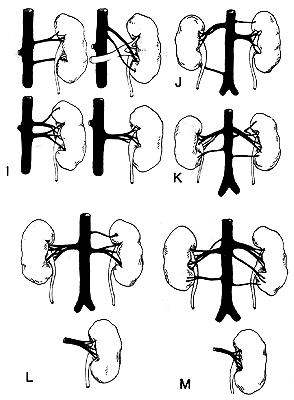
I: Upper left, superior polar artery arising from one or two main renals. Upper right, accessory artery to lower pole crossing, renal vein obliquely. This specimen shows how difficult it would be to secure the bleeding trunk if such an artery were over looked. Lower left, upper of two main renal arteries gives off a superior polar artery, and a retropelvic artery. Lower right, main renal artery divides into pre- and retropelvic branches of equal size.
J: On the right side is seen a typical division of the main renal into pre- and retropelvic branches of equal size. Note lower polar accessory artery to right kidney lying behind ureter. There is also an accessory superior polar on the left side.
K: Anterior view showing retropelvic artery on right side arising from renal, close to aorta. On the left side note the superior polar and the retropelvic artery, both arising from one of the two main renals. Note inferior polar (accessory) arteries arising from aorta on both sides.
L: Upper, right kidney. Typical example on right side of division of main renal artery into equal-sized pre- and retropelvic branches. Lower, example of fan-like division of retropelvic artery over posterior aspect of pelvis.
M: Upper, anterior view. Right kidney shows a single accessory artery to lower pole. Left kidney shows two accessory arteries to lower pole, all from aorta. Lower, posterior view of right kidney showing typical fan-like distribution of retropelvic artery.
Redrawn from Eisendrath, D.N. The relations of variations in the renal vessels to pyelotomy and nephrectomy. Ann. Surg. 71:726-743, 1920.
Section Top | Title Page
Please send us comments by filling out our Comment Form.
All contents copyright © 1995-2024 the Author(s) and Michael P. D'Alessandro, M.D. All rights reserved.
"Anatomy Atlases", the Anatomy Atlases logo, and "A digital library of anatomy information" are all Trademarks of Michael P. D'Alessandro, M.D.
Anatomy Atlases is funded in whole by Michael P. D'Alessandro, M.D. Advertising is not accepted.
Your personal information remains confidential and is not sold, leased, or given to any third party be they reliable or not.
The information contained in Anatomy Atlases is not a substitute for the medical care and advice of your physician. There may be variations in treatment that your physician may recommend based on individual facts and circumstances.
URL: http://www.anatomyatlases.org/