
Illustrated Encyclopedia of Human Anatomic Variation: Opus II: Cardiovascular System
Ronald A. Bergman, PhD
Adel K. Afifi, MD, MS
Ryosuke Miyauchi, MD
Peer Review Status: Internally Peer Reviewed

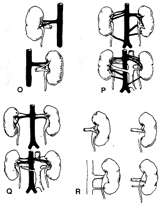
O: Upper, anterior view. Lower, posterior view of right kidney showing main renal vein crossing posterior aspect of pelvis.
P: Right kidney. Upper, Accessory upper polar artery arising close to aorta from one of two main renal arteries. Note also accessory lower polar artery arising from aorta close to common iliac. Similar condition in left kidney but superior polar artery arises from one of two main renals close to kidney. Lower, posterior view of upper figure, showing a large renal vein and artery entering kidney back of pelvis on left side, and a smaller retropelvic vein directly from the vena cava.
Q: Upper, front view showing superior polar artery arising from main renal on right side and from aorta on left side. Lower, posterior view of upper figure, showing a fairly large renal vein crossing middle of back of pelvis.
R: Diagrammatic representation of various types of retropelvic veins: Upper left, division of the main renal vein into pre- and retropelvic branches of equal size; upper right, main renal vein crosses posterior aspect of pelvis to reach kidney; lower left, accessory renal vein to lower pole of kidney; lower right, main renal vein divides into two branches, the larger of which is retropelvic. There is also an accessory renal vein directly from aorta.
Eisendrath provided the following practical conclusions from all published statistics.
"It is of importance for the surgeon to remember that the examination of 1237 kidneys by various investigators reveals the fact that upper polars from the renals occurred in 68, or 16 percent, of 518 kidneys. Upper polars from the aorta were found in 68, or .5 percent, and lower polars from the aorta in 71, or nearly .6 percent, of 1237 kidneys. Lower polars from the iliacs were found in only 6, or .04 percent, of the 1237 kidneys. "
"According to my Eisehdrath's present series of dissections, one can expect to find upper polar arteries arising from the main renals in about one kidney out of five. Upper polars arising from the aorta were found in one out of about 17 kidneys and lower polars (from the aorta and iliacs) in one kidney out of about 7 kidneys.
Adding together the observations of all previous investigators and our own we find that (a) upper polars arising from the main renals occur in about one out of 200 kidneys; (b) upper polars arising from the aorta in about one out of about 190 kidneys; and (c) lower polars from the main renal, the aorta, or common iliacs in one out of about 185 kidneys. Although accessory polar vessels did not occur as frequently as stated by Quain, i.e., 20 percent, they are found often enough to be constantly borne in mind during operation."
The Retropelvic Vessels
"The tradition still exists that one needs only to guard against
injury of a retropelvic artery which pursues a more or less typical
course in the sinuses formed at the point where the kidney tissue
slightly overlaps the renal pelvis. That there may be (a) variations
from this arch-like distribution of the artery and (b) that one or
more large veins, even the main renal, may cross our field of
operations are two anatomical facts which deserve more widespread
knowledge in order to avoid injury to these anomalous vessels duiring
pyelotomy.
In Albarran's book, published in 1910, reference is made to retropelvic artery and vein to the effect that the main renal artery may divide into pre- and retropelvic trunks of equal size, and that the retropelvic artery on its way to the sinus gives off branches similar to those arising from the prepelvic artery. His only statement in regard to the retropelvic vein is that it is not constant - was found in 5 of 29 cases by Hauch - and finally that it may prove a source of trouble during pyelotomy.
In view of the results of my present dissections, I believe we must abandon the view that the posterior aspect of the renal pelvis is the avascular field which we have generally believed it to be. The distribution of the prepelvic vessels seldom, if ever, enters into consideration in the operation of pyelotomy, because the route of election is through the less vascular field.
In a total of 218 kidneys, the following observations of variations of the retropelvic vessels of surgical importance were made:
A. Anomalies of the Retropelvic Artery Alone. 1 . Division of the single main renal artery into equal-sized branches was found very frequently.
2. When there were two main renal arteries, one of these frequently became the retropelvic, i.e., the latter arose directly from the aorta instead of the main renal artery. This was found in 5 kidneys out of 124.
3. The main renal or one of two renal arteries was found to be retropelvic in 2 kidneys out of 124.
4. The retropelvic artery had its origin frorn an accessory lower polar artery in 2 kidneys out of 124.
5. A retropelvic artery directly from the aorta was found in 2 kidneys out of 124.
6. The main retropelvic artery does not cling in an arch-like manner to the renal sinus. This generally accepted course of the vessel may be described as the high type to distinguish it from various combinations which were found in both the Illinois and Harvard dissections. These extra vessels which may give rise to troublesome bleeding during pyelotomy are (a) a high middle and low or fan-like distribution found twice in 124 kidneys; (b) a high and middle type of branching found twice in 124 kidneys; (c) a high and low type found twice in 124 kidneys; (d) a single artery crossing the middle of the pelvis, found four times in 124 kidneys; and finally (e) a middle and low type found seven times in 124 kidneys.
B. Anomalies of the Retropelvic Veins. 1. One large vein arising from the vena cava was found five times in 218 kidneys, passing directly across the back of the renal pelvis.
2. The main renal vein divided into equal-sized pre- and retropelvic branches in 3 of 218 kidneys. The retropelvic branch passed directly across the pelvis and, as in the case of the preceding anomaly, could be easily injured during pyelotomy.
3. The most important anomaly, so far as the veins were concerned, was that the main renal vein, instead of being prepelvic, was retropelvic in 9 out of 218 kidneys.
C. Anomalies Involving Both Retropelvic Vein and Artery. 1. One large vein directly from the vena cava and one artery directly from the aorta crossed the back of the pelvis in one of 94 kidneys.
2. Two large veins directly from the vena cava and one artery from the aorta crossed the back of the pelvis in one of 94 kidneys."
Types of Pelvis
Observations made as to the relative frequency of the various, types
of renal pelvis revealed the following:
1. The single or ampullary pelvis was found in 84 (89 percent) out of 94 kidneys.
2. The divided or bifid type was found in 7 (8 percent) out of 94 kidneys. In 4 of these it was present on both sides.
3. The trifid type was found in 3 percent of 94 kidneys.
Authors' note: Eisendrath's references to a book by Albarran (1910) and to the work of Hauch were not found in his bibliography.
Redrawn from Eisendrath, D.N. The relations of variations in the renal vessels to pyelotomy and nephrectomy. Ann. Surg. 71:726-743, 1920.
Section Top | Title Page
Please send us comments by filling out our Comment Form.
All contents copyright © 1995-2024 the Author(s) and Michael P. D'Alessandro, M.D. All rights reserved.
"Anatomy Atlases", the Anatomy Atlases logo, and "A digital library of anatomy information" are all Trademarks of Michael P. D'Alessandro, M.D.
Anatomy Atlases is funded in whole by Michael P. D'Alessandro, M.D. Advertising is not accepted.
Your personal information remains confidential and is not sold, leased, or given to any third party be they reliable or not.
The information contained in Anatomy Atlases is not a substitute for the medical care and advice of your physician. There may be variations in treatment that your physician may recommend based on individual facts and circumstances.
URL: http://www.anatomyatlases.org/