
Illustrated Encyclopedia of Human Anatomic Variation: Opus II: Cardiovascular System
Ronald A. Bergman, PhD
Adel K. Afifi, MD, MS
Ryosuke Miyauchi, MD
Peer Review Status: Internally Peer Reviewed

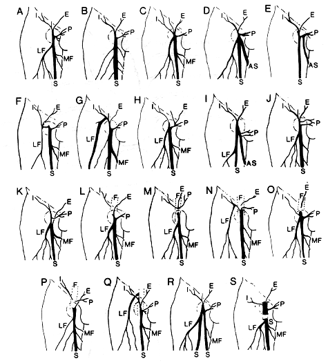
I, Superficial circumflex iliac vein; E, superficial epigastric vein; P, superficial external pudendal vein; LF, lateral superficial femoral vein; MF, medial and superficial femoral vein; AS, accessory saphenous vein; SS, double saphenous vein; S, vena saphena magna; F, femoral vein.
A: Average "textbook" diagram of venous drainage at fossa ovalis. Incidence, 37%.
B: Multiple divisions of the medial and lateral femoral veins of small caliber. Incidence, 6%.
C: The large lateral superficial femoral vein drains into the fossa ovalis. The inconstant thoracoepigastric vein drains into the vena saphena magna instead of into the femoral vein. Incidence, 2%.
D: The lateral superficial femoral and the accessory saphenous vein drain into the fossa ovalis. Incidence, 2%.
E: The accessory saphenous vein forms a common stem with the superficial external pudendal vein before joining the vena saphena magna. Incidence, 6%.
F: A common trunk formed by the lateral superficial femoral, superficial circumflex iliac, and superficial epigastric veins drains into the fossa ovalis. Incidence, 9%.
G: A common trunk formed by the lateral superficial femoral and the superficial circumflex iliac vein drains into the fossa ovalis. Incidence, 9%.
H: The superficial epigastric and the superficial external pudendal vein form a common trunk. A large lateral superficial femoral vein is present. Incidence, 2%.
I: An accessory saphenous vein is present. Note the drainage of double superficial external pudendal veins. Incidence, 1%.
J: Double superficial external pudendal veins drain into the fossa ovalis. Incidence, 3%.
K: The superficial epigastric vein drains into the vena saphena magna below the fossa ovalis. Incidence, 3%.
L: The superficial circumflex iliac vein drains into the femoral vein. Incidence, 1%.
M: All high collateral veins drain directly into the femoral vein. Incidence, 6%.
N: The lateral femoral and the superficial circumflex iliac vein form a common trunk. The other high collateral veins drain directly into the femoral vein. Incidence, 1%.
O: The lateral femoral vein drains into the fossa ovalis. The superficial epigastric vein drains directly into the femoral vein. Incidence, 6%.
P: Note the small caliber multiple medial and lateral superficial femoral veins. The superficial circumflex iliac and the superficial external pudendal veins drain directly into the femoral vein. Incidence, 1%.
Q: The lateral superficial femoral vein drains directly into the femoral vein. Incidence, 1%.
R: A double vena saphena magna with joining at the fossa ovalis. Incidence, 3%.
S: The saphena magna pierces the deep fascia to enter the femoral vein about 1 inch below the fossa ovalis. Incidence, 1%.
Redrawn from Glasser, S.T An anatomic study of venous variations at the fossa ovalis. Arch. Surg. 46:289-295, 1943.
Section Top | Title Page
Please send us comments by filling out our Comment Form.
All contents copyright © 1995-2024 the Author(s) and Michael P. D'Alessandro, M.D. All rights reserved.
"Anatomy Atlases", the Anatomy Atlases logo, and "A digital library of anatomy information" are all Trademarks of Michael P. D'Alessandro, M.D.
Anatomy Atlases is funded in whole by Michael P. D'Alessandro, M.D. Advertising is not accepted.
Your personal information remains confidential and is not sold, leased, or given to any third party be they reliable or not.
The information contained in Anatomy Atlases is not a substitute for the medical care and advice of your physician. There may be variations in treatment that your physician may recommend based on individual facts and circumstances.
URL: http://www.anatomyatlases.org/