
Illustrated Encyclopedia of Human Anatomic Variation: Opus II: Cardiovascular System
Ronald A. Bergman, PhD
Adel K. Afifi, MD, MS
Ryosuke Miyauchi, MD
Peer Review Status: Internally Peer Reviewed
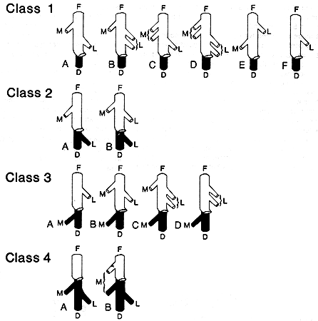
D, Deep femoral vein (shaded black); F, femoral vein; L, lateral femoral circumflex vein; M, medial femoral circumflex vein.
The deep femoral vein empties into the femoral vein about 8 cm distal to the inguinal ligament.
The authors make the following observations based on their study (the classes are here referred to as I, II, III, and IV):
Observations
" It was found by Charles et al. [reference 226] that the
modes of termination might be placed in one of four classes, and the
same method of classification has been followed in this paper
although the number of types has increased with the increase in the
number of observations.
Classes.
Class I consists of those cases in which the circumflex veins
terminate directly in the femoral vein [see figure] * In
class II the lateral circumflex vein terminates in the deep femoral,
while the medial circumflex vein ends directly in the femoral. The
reverse is true of class III, the medial circumflex terminating in
the deep femoral, and the lateral circumflex vein emptying into the
femoral itself. In class IV both the medial and lateral circumflex
veins terminate in the deep femoral vein.
Types.
The several types composing each class permit classification as to
the relative position, duplicity or absence of one or other of the
circumflex veins.
Discussion
It was noted in an accompanying table I [not provided here]
that class I, in which the circumflex veins terminate directly in the
femoral, includes 86.14 per cent of the 541 observations. This is in
contradiction to the statements made in textbooks which describe the
circumflex veins as terminating in the deep femoral (our class IV).
In the present series, only 1.85 per cent of the veins ended in that
manner.
Of the six types making up class I, type IA constitutes 64.87 per cent of all observations. As is shown in the diagram, there are in this type single medial and lateral circumflex veins, both terminating in the femoral, the medial circumflex vein being the more proximal.
Next in frequency is type IB (12.57 per cent), which is quite similar to IA with the exception that there are two lateral circumflex veins distal to a single medial circumflex vein, all of which empty directly into the femoral. Type IC (3.70 per cent) has two medial circumflex veins proximal to a single lateral circumflex vein.
In all three of the above types (IA, IB, and IC) the termination of the medial circumflex vein or veins is proximal to that of the lateral circumflex vein. Since these three types together make up 81.14 per cent of all the cases observed, it may be concluded that in a large majority of the cases the termination of the lateral circumflex is distal to that of the medial circumflex vein. Type ID is an interesting example of this tendency. In this type there are two medial circumflex veins and two lateral circumflex veins and although the two medial circumflex veins are not both proximal to the two lateral circumflex veins, each medial circumflex is proximal to its corresponding lateral circumflex. In addition, in classes other than class I, where the circumflex veins do not characteristically end in the femoral, there is still a tendency for the medial circumflex to end above the lateral circumflex.
In type IE (2.96 per cent) we find for the first time a change in this characteristic relationship of the medial and lateral circumflex veins, the lateral circumflex being proximal to the medial circumflex. In type IF (0.92 per cent) the medial circumflex vein is absent.
Classes II and III constitute together but 11.46 per cent of the observations, class III predominating slightly (6.28 per cent as compared with 5.18 per cent).
Class II is made up of two types, the first and predominating (IIA, 4.07 per cent) having a single medial circumflex emptying into the femoral, and a single lateral circumflex emptying into the deep femoral. Type IIB (1.11 per cent) differs from IIA only in having an accessory lateral circumflex ending in the femoral, just distal to the medial circumflex. Here again in both these types the position of the medial circumflex is characteristically proximal to that of the lateral circumflex vein.
There are four types making up class III, type IIIA being found most frequently (3.51 per cent). This type has a single medial circumflex terminating in the deep femoral, while the lateral circumflex terminates superiorly in the femoral. Type IIIB differs from IIIA only in having an accessory medial circumflex emptying into the femoral itself, immediately proximal to the lateral circumflex vein. Again there is but a small variation that distinguishes IIIB from IIIC, namely, the presence of two adjacent lateral circumflex veins rather than a single vein. Type IIID has duplicate lateral circumflex veins emptying into the femoral and a single medial circumflex emptying into the deep femoral. These two last-named types are only rarely found, representing but 0.74 per cent and 0.37 per cent of the present series, respectively.
As mentioned above, in only 1.85 per cent of the cases do both the lateral and medial circumflex veins end in the deep femoral (class IV). The most common type in this class is type IVA which has a single medial circumflex and a single lateral circumflex, both emptying into the deep femoral. The other type IVB, has an accessory medial circumflex ending in the femoral, and was found in only one case."
Redrawn from Baird, R.D. and J.S. Cope. On the termination of the circumflex veins of the thigh and their relations to the origins of the circumflex arteries. Anat Rec. 57:325-337, 1933.
Section Top | Title Page
Please send us comments by filling out our Comment Form.
All contents copyright © 1995-2024 the Author(s) and Michael P. D'Alessandro, M.D. All rights reserved.
"Anatomy Atlases", the Anatomy Atlases logo, and "A digital library of anatomy information" are all Trademarks of Michael P. D'Alessandro, M.D.
Anatomy Atlases is funded in whole by Michael P. D'Alessandro, M.D. Advertising is not accepted.
Your personal information remains confidential and is not sold, leased, or given to any third party be they reliable or not.
The information contained in Anatomy Atlases is not a substitute for the medical care and advice of your physician. There may be variations in treatment that your physician may recommend based on individual facts and circumstances.
URL: http://www.anatomyatlases.org/