
Illustrated Encyclopedia of Human Anatomic Variation: Opus II: Cardiovascular System
Ronald A. Bergman, PhD
Adel K. Afifi, MD, MS
Ryosuke Miyauchi, MD
Peer Review Status: Internally Peer Reviewed
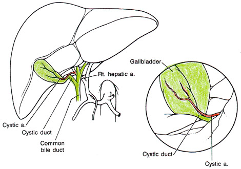
Figure 1: The "normal" or most frequent pattern, shown on the left, consists of a single cystic artery that runs through Calot's triangle (the space between the hepatic duct, cystic duct, and undersurface of the liver) to enter the left or anterior surface of the gallbladder. Note the superficial branch of the cystic artery, visible on the surface of the gallbladder, which provides a useful visual landmark leading to the cystic artery. The laproscopic appearance is show in the inset view on the right. The cystic duct is closer to the laporscope and appears larger than the cystic artery, which is smaller and further away. The superficial cystic artery is again visible on the surface of the gallbladder. This normal pattern is variably reported as being present in 35% to 75% of individuals [2-6]. a.=artery; Rt.=right.
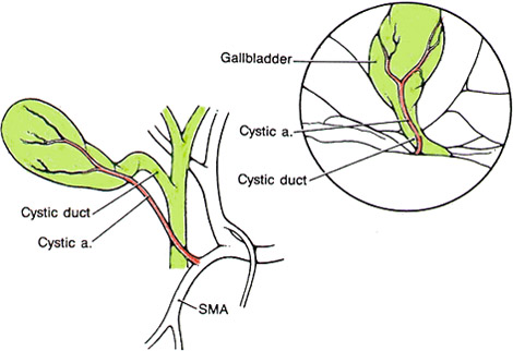
Figure 2:In this example, a low-lying or posterior cystic artery is shown arising from the superior mesenteric artery (SMA) and making an early ascent to the gallbladder. Hence, it lies closer to the laporoscope than the cystic duct and may appear in the position normally occupied by the cystic duct (inset). This transposed cystic artery-cystic duct relationship may occur in as many as 18% of individuals [4] and is the most common laparoscopic arterial anomaly reported [1]. A single aberrant cystic artery is present in 4% to 12% [1,7,9]. Double cystic arteries are more common, occurring in around 16% of individuals [3,6]. a.=artery.
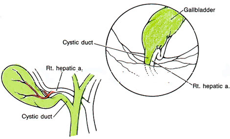
Figure 3: A tortuous or caterpillar hump right hepatic artery appears in the position of the cystic artery, but it is larger than the normal cystic artery. This anomaly occurs in 4% to 15% of patients [1-3,5]. The unusually large size of the vessel provides an important clue to the laparoscopic surgeon (inset). a.=artery; Rt.=right.
from Scott-Conner.
Section Top | Title Page
Please send us comments by filling out our Comment Form.
All contents copyright © 1995-2024 the Author(s) and Michael P. D'Alessandro, M.D. All rights reserved.
"Anatomy Atlases", the Anatomy Atlases logo, and "A digital library of anatomy information" are all Trademarks of Michael P. D'Alessandro, M.D.
Anatomy Atlases is funded in whole by Michael P. D'Alessandro, M.D. Advertising is not accepted.
Your personal information remains confidential and is not sold, leased, or given to any third party be they reliable or not.
The information contained in Anatomy Atlases is not a substitute for the medical care and advice of your physician. There may be variations in treatment that your physician may recommend based on individual facts and circumstances.
URL: http://www.anatomyatlases.org/