
Illustrated Encyclopedia of Human Anatomic Variation: Opus III: Nervous System
Ronald A. Bergman, PhD
Adel K. Afifi, MD, MS
Ryosuke Miyauchi, MD
Peer Review Status: Internally Peer Reviewed
In many older textbooks of anatomy there is a brief but accurate description of these two cavities. Dandy describes these as follows:
"The nomenclature, however, is not uniform. For example, the cavum septi pellucidi is perhaps better known as the fifth ventricle, and the cavum vergae is called Verga's ventricle, the sixth ventricle, the ventricle of Strambio, ventriculus fornicis, ventriculus triangularis, and the canal aqueduct.
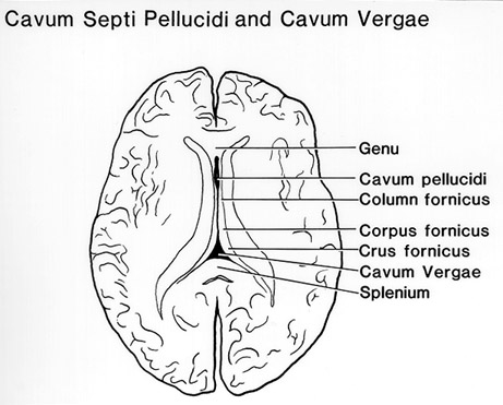
Position and Boundaries of the Cavities
The corpus callosum defines the anterior, superior and posterior limits of the two cavities which, when not continuous, are separated from each other by the anterior limit of the fornix as it courses obliquely backward and upward from the anterior comissure to the body of the corpus callosum. Being of congenital origin, the two cavities are doubtless dependent on the development of the corpus callosum and the fornix. They may coexist and be isolated from each other when the fornix is intact; they may coexist and be in communication through a defect in the fornix, or they may form a single large cavity when the fornix is not attached to the corpus callosum. The cavum septi pellucidi is frequently present when the cavum vergae is absent, and Verga's cavity may be present when the cavum septi pellucidi is absent. Kauffmann stated that the cavum vergae may exist on one side of the midline and be absent on the other.
The cavum septi pellucidi has the following boundaries: anteriorly, the genu of the corpus callosum; superiorly, the body of the corpus callosum; corpus callosum, posteriorly, the anterior limb and pillars of the fornix, inferiorly, the rostrum of the corpus callosum and the anterior commissure, laterally, the layers of the septum pellucidum. Viewed laterally, the cavum septi pellucidi is roughly triangular with the base at the corpus callosum. Viewed in cross-section the cavity is also triangular with the base at the corpus callosum.
The cavum vergae has the following boundaries anteriorly, the anterior limb of the fornix, superiorly, the body of the corpus callosum, posteriorly, the splenium of the corpus callosum, inferiorly, the psalterium (lyra davidis) and hippocampal commissure, the fibers of which bridge the space between the diverging posterior pillars of the fornix. This cavity is also triangular when viewed from the side. The cavum vergae flares out laterally on both sides with the curve of the fornix and pushes under the lateral ventricles at its extreme lateral extensions.
In most adult brains both spaces are absent or are at most potential, but in every 100 necropsies actual cavities of varying size will be seen. Neither cavity can be regarded as part of the great ventricular system in which cerebrospinal fluid forms and through which it circulates."
From Dandy, W.E. Congenital cerebral cysts of the cavum septi pellucidi (fifth ventricle) and cavum vergae (sixth ventricle). Diagnosis and treatment. Arch. Neurol Psychiatry 25:44-66, 1931.
Section Top | Title Page
Please send us comments by filling out our Comment Form.
All contents copyright © 1995-2024 the Author(s) and Michael P. D'Alessandro, M.D. All rights reserved.
"Anatomy Atlases", the Anatomy Atlases logo, and "A digital library of anatomy information" are all Trademarks of Michael P. D'Alessandro, M.D.
Anatomy Atlases is funded in whole by Michael P. D'Alessandro, M.D. Advertising is not accepted.
Your personal information remains confidential and is not sold, leased, or given to any third party be they reliable or not.
The information contained in Anatomy Atlases is not a substitute for the medical care and advice of your physician. There may be variations in treatment that your physician may recommend based on individual facts and circumstances.
URL: http://www.anatomyatlases.org/