
Illustrated Encyclopedia of Human Anatomic Variation: Opus III: Nervous System
Ronald A. Bergman, PhD
Adel K. Afifi, MD, MS
Ryosuke Miyauchi, MD
Peer Review Status: Internally Peer Reviewed
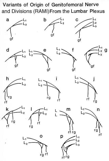
In this study of 200 bodies (400 nerves), 80.2% presented as a single trunk and 19.75% as two rami: genital and femoral. The trunk or rami were usually formed by nerve fibers from L1, L2 from the lumbar plexus. The level of emergence of the nerve or nerves from the psoas major muscle and the level of division into terminal branches were individually highly variable. These characteristics were unrelated to each other, sex, age, height or weight or side of the body. The genitofemoral nerve was present in all cases. Two types were recognized. In type I, the trunk of the nerve divided into two main rami: genital and femoral. In type II, the rami arose separately from the lumbar plexus. Type I was present bilaterally in 70.5%, and type II in 10% of cases.
The manner in which the genitofemoral nerve arose from the lumbar plexus varied markedly. Nerves of type I arose from a single root in 75.5%, and from two roots in 4.5% of cases. Single root nerves arose by union of the ventral branches of spinal nerves L1, L2 (A) in 75%, from L2 (B) in 0.5%, and by union of L2, L3 (C) in 0.25% of cases. In double-rooted nerves (C-G) the upper root arose from L, in 3.75%, and from L1, L2 in 0.75% of cases; and the lower root from L1, L2 in 1.75%, from L2 in 2.5%, and from L3 in 0.25%. Both roots, after a short Course, joined within the substance of the psoas major muscle. In type II, three variations of origin of the nerve from the lumbar plexus were observed. In the first variant, present in 18.25% of cases, both the genital and femoral branch arose from the plexus by one root. Both branches were derived from L1, L2 in 17.25% of cases (H); the genital branch from L, and the femoral branch from L1, L2 (1) in 0.5%; and the genital branch from L1, L2 and the femoral branch from L, (J) in 0.5% of cases. In the second variant, present in 1.0% of cases, the genital branch arose by one root, and the femoral branch by two (K-N). In three cases the genital branch arose from L1, L2, and in one case from L1. The upper root of the femoral branch in three cases arose from L1, in one case from L1, L2; and the lower root in three cases arose from L1, L2, and in one case from L2. After a short course, the two roots of the femoral branch united within the psoas major muscle. In the third variant, represented in 0.5% of cases, the genital branch had two roots, and the femoral branch had one (0, P). The upper root of the genital branch arose from L, or L1, L2, and the lower root from L1, L2, or L3. Both roots united just after leaving the plexus. The femoral branch in this variant was formed from L1, L2, In type II of the genitofemoral nerve, the genital branch arose from the plexus above the femoral branch in 18.5%, and from the femoral branch in 1.25% of cases.
The origin of the genitofemoral nerve from the lumbar plexus was symmetric in 64% of cases in type I, and in 7.5% in type II. Spinal nerve L1, was present in nearly all cases, and L2 in all cases sent fibers to the genitofemoral nerve. Fibers from L3 were present in only 0.75% of cases.
L1, to L.3, respective lumbar nerves; gf, genitofemoral nerve; rf, femoral branch; rg, genital branch. Redrawn from Urbanowicz, Z. External structure of the genitofemoral nerve in postfetal life in man. Folia Morphol. (Warsaw) 34:425-435, 1975.Section Top | Title Page
Please send us comments by filling out our Comment Form.
All contents copyright © 1995-2024 the Author(s) and Michael P. D'Alessandro, M.D. All rights reserved.
"Anatomy Atlases", the Anatomy Atlases logo, and "A digital library of anatomy information" are all Trademarks of Michael P. D'Alessandro, M.D.
Anatomy Atlases is funded in whole by Michael P. D'Alessandro, M.D. Advertising is not accepted.
Your personal information remains confidential and is not sold, leased, or given to any third party be they reliable or not.
The information contained in Anatomy Atlases is not a substitute for the medical care and advice of your physician. There may be variations in treatment that your physician may recommend based on individual facts and circumstances.
URL: http://www.anatomyatlases.org/