
Illustrated Encyclopedia of Human Anatomic Variation: Opus III: Nervous System
Ronald A. Bergman, PhD
Adel K. Afifi, MD, MS
Ryosuke Miyauchi, MD
Peer Review Status: Internally Peer Reviewed
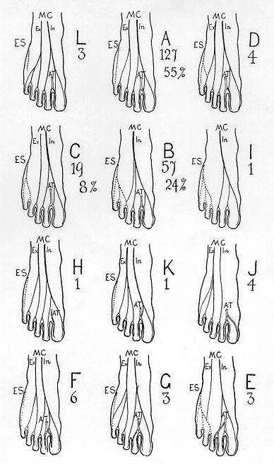 "A total of 229 feet have been examined, and in tabulating
the results the graphic method has been adopted in preference to
descriptive. Twelve types are figured, and lettered A, B, C, &
c.., in order of their frequency. The first six are arranged in such
a way as to show how the external saphenous replaces the external
division of the musculo-cutaneous nerve, or vice versa. The last six
(H, K, I, F, G, E) are arranged to display the progressive variations
in the cutaneous distribution of the anterior tibial nerve. In these
types connecting loops between the nerves have in some cases omitted,
as the information necessary was sufficiently complete. The figures
attached to the types indicate the numbers of such specimens out of
the total of 229 examined. In some cases the percentage is also
given.
The musculo-cutaneous is represented by a solid black line;
the external saphenous by a dotted line. The anterior tibial by a
double outline.
The so-called normal arrangement represented by type A
occurs in 55 percent of cases. Next in order is type B, where the
external saphenous supplies the two and a half outer toes. This
distributon of the nerves was met with in 24 percent of the feet
examined.
In one case, the internal saphenous supplied the inner side
of the great toe; in another, it reached the inner side of the head
of the metatarsal bone."
AT, Anterior tibial (deep peroneal) nerve; ES, external
saphenous (sural) nerve; MC, musculocutaneous (superficial peroneal)
nerve: Ex, external (lateral) branch; and In, internal (medial)
branch.
From Thomson, A. Second annual report of the committee of
collective investigation of the Anatomical Society of Great Britain
and Ireland for the year 1890-91. J. Anat. Physiol. 26:76-93, 1891.
"A total of 229 feet have been examined, and in tabulating
the results the graphic method has been adopted in preference to
descriptive. Twelve types are figured, and lettered A, B, C, &
c.., in order of their frequency. The first six are arranged in such
a way as to show how the external saphenous replaces the external
division of the musculo-cutaneous nerve, or vice versa. The last six
(H, K, I, F, G, E) are arranged to display the progressive variations
in the cutaneous distribution of the anterior tibial nerve. In these
types connecting loops between the nerves have in some cases omitted,
as the information necessary was sufficiently complete. The figures
attached to the types indicate the numbers of such specimens out of
the total of 229 examined. In some cases the percentage is also
given.
The musculo-cutaneous is represented by a solid black line;
the external saphenous by a dotted line. The anterior tibial by a
double outline.
The so-called normal arrangement represented by type A
occurs in 55 percent of cases. Next in order is type B, where the
external saphenous supplies the two and a half outer toes. This
distributon of the nerves was met with in 24 percent of the feet
examined.
In one case, the internal saphenous supplied the inner side
of the great toe; in another, it reached the inner side of the head
of the metatarsal bone."
AT, Anterior tibial (deep peroneal) nerve; ES, external
saphenous (sural) nerve; MC, musculocutaneous (superficial peroneal)
nerve: Ex, external (lateral) branch; and In, internal (medial)
branch.
From Thomson, A. Second annual report of the committee of
collective investigation of the Anatomical Society of Great Britain
and Ireland for the year 1890-91. J. Anat. Physiol. 26:76-93, 1891.
Section Top | Title Page
Please send us comments by filling out our Comment Form.
All contents copyright © 1995-2024 the Author(s) and Michael P. D'Alessandro, M.D. All rights reserved.
"Anatomy Atlases", the Anatomy Atlases logo, and "A digital library of anatomy information" are all Trademarks of Michael P. D'Alessandro, M.D.
Anatomy Atlases is funded in whole by Michael P. D'Alessandro, M.D. Advertising is not accepted.
Your personal information remains confidential and is not sold, leased, or given to any third party be they reliable or not.
The information contained in Anatomy Atlases is not a substitute for the medical care and advice of your physician. There may be variations in treatment that your physician may recommend based on individual facts and circumstances.
URL: http://www.anatomyatlases.org/