
Illustrated Encyclopedia of Human Anatomic Variation: Opus IV: Organ Systems
Ronald A. Bergman, PhD
Adel K. Afifi, MD, MS
Ryosuke Miyauchi, MD
Peer Review Status: Internally Peer Reviewed
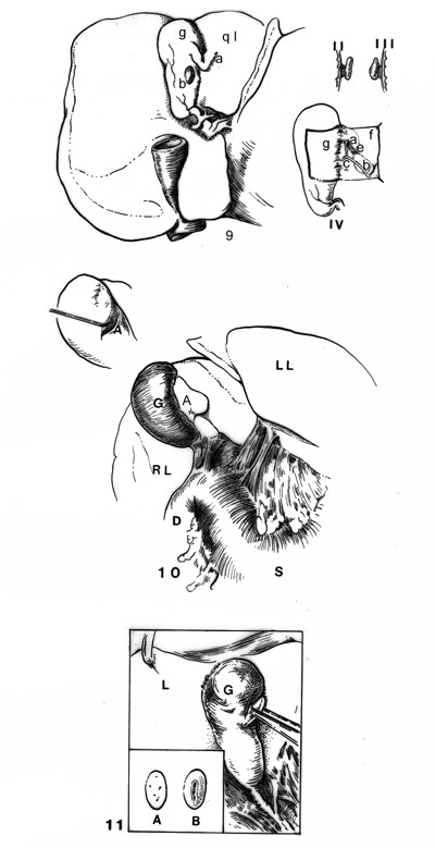
Fig. 9 - Accessory lobe of the liver springing from undersurface of gallbladder. This is probably the first picture of such as condition; it appeared in the Marseille médical 59:442, 1922. I, b, a small accessory lobe attached to surface of gallbladder. Passing from the quadrate lobe (ql) to the gallbladder is V or crochet-shaped piece of liver tissue, a. It is definitely separated from the accessory lobe. II, accessory lobe projecting from gallbladder. It is attached by a definite mesentery. III, accessory lobe viewed from the left side. IV, peritoneum, f, of gallbladder has been dissected free and reflected to the side; a, the V-shaped piece of liver tissue passing from the quadrate lobe to the gallbladder; b, the accessory lobe. Passing from the accessory lobe, b, to the V-shaped piece of liver tissue, a, are: c, the arteriole, d, the vein, and e, the biliary duct; all undoubtedly in this way reached the liver proper; gis the musculature of the gallbladder. This picture has been redrawn by Max Brödel and turned upside down in order that its relations might be compared with our case. (After Frederic Corsy.)
Fig 10- Accessory lobe springing from quadrate lobe and intimately blended with left surface of gallbladder. This specimen was found in the dissecting room. The body was that of a colored woman, aged 55. The liver showed a slight tendency to lobulation, as noted in the two fissures on the under surface of the left lobe. The accessory lobe was rather flat and somewhat triangular and had rounded edges. In over half its extent it was intimately connected with the gallbladder and liver, as shown in the sketch in the upper left corner of the picture. The lower half of the accessory lobe was free. A triangular peritoneal fold passed from the lower border of the lobe to the gallbladder. There were adhesions from the stomach to the liver and from duodenum to the gallbladder.
Fig. 11- Accessory lobe of liver springing from surface of gallbladder (G.) (actual size). The undersurface of the liver is shown. The gallbladder looks normal but at operation in its lower portion it was found loosely attached to the duodenum. Attached to the gallbladder by a short mesentery is a miniature liver. It was lifted up, the pedicle was tied with a catgut ligature, and removed. It measured 12 by 7 by 3.5 mm. in its central portion: ashows the smooth surface of the accessory liver, bits under surface. Traversing the pedicle were a branch of the hepatic artery, a branch of the hepatic vein, a branch of the portal vein, and a branch of the hepatic duct. The pedicle at the level seen in the picture shows a longitudinal section of what appears to be a branch of the protal vein. L. liver.
Section Top | Title Page
Please send us comments by filling out our Comment Form.
All contents copyright © 1995-2024 the Author(s) and Michael P. D'Alessandro, M.D. All rights reserved.
"Anatomy Atlases", the Anatomy Atlases logo, and "A digital library of anatomy information" are all Trademarks of Michael P. D'Alessandro, M.D.
Anatomy Atlases is funded in whole by Michael P. D'Alessandro, M.D. Advertising is not accepted.
Your personal information remains confidential and is not sold, leased, or given to any third party be they reliable or not.
The information contained in Anatomy Atlases is not a substitute for the medical care and advice of your physician. There may be variations in treatment that your physician may recommend based on individual facts and circumstances.
URL: http://www.anatomyatlases.org/