
Illustrated Encyclopedia of Human Anatomic Variation: Part V: Skeletal Systems
Ronald A. Bergman, PhD
Adel K. Afifi, MD, MS
Ryosuke Miyauchi, MD
Peer Review Status: Internally Peer Reviewed
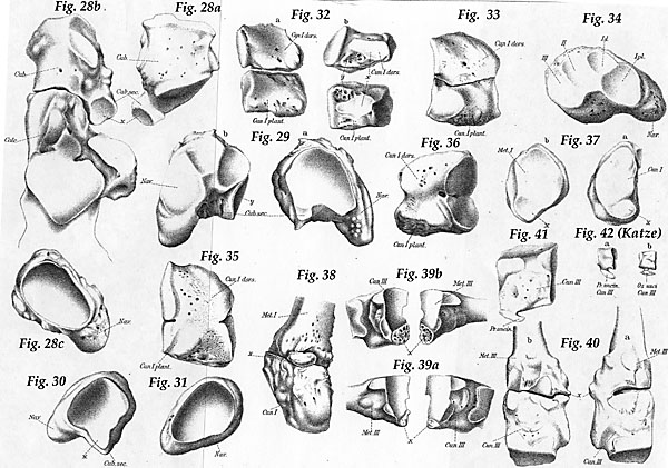
Figure 28:Cuboides secondarium.
a:Right cuboid with cuboides secondarium; x = Joint surface for the head of the talis.b:Calcaneus (calc.) and cuboid (cub.) in-situ, cauboides secondarius with fused cuboid; x = joint surface of the cuboid secondarius for the head of the talus.
c: Left naviculare without cuboid.
Figure 29:Cuboides secondarium. Fused with the navicular.
a:Proximal surface; x = joint surface for the head of the talus.b: Distal surface.
Figure 30:Right navicualr with cuboides secondarium. x = special facette of the head of the talus. Cub. sec. = cuboides secondarium; Nav. = navicular.
Figure 31:Right navicular without cuboides secondarium. The latter is fused with the cuboid. Nav. = navicular.
Figure 32:Bipartite cuneiform I.
a:Both pieces in-situ.b: The contact surface; partly joint surface, partly the coalescent surface. Cun. I dors. = dorsal surface cuneiform I.; cun I plant. = plantar surface of cuneiform I.
Figure 33:Bipartite cuneiform I. Medial surface.
Figure 34:Navicualr with four wedge-shaped facets, with the first two joined. Id. = Facet for the dorsal half; I pl = Facet for the plantar half.
Figure 35:Cuneiform I imperfectly bipartite. Medial surface.
Figure 36:Cuneiform I imperfectly bipartite. Medial surface.
Figure 37:Special lower facet in the tarso-metatarsal joint.
a:Distal surface of cuneiform Ib:Proximal surface of metatarsal I; x = lower facet.
Figure 38: Pars peronea metatarsal I. Fibulo-plantar view. x = Pars peronea.
Figure 39:Coalescence of cuneiform-metatarsal III.
a:Right weak.b:Left strong; coalescence surface. Tibial surface.
Figure 40:Coalescence of cuneiform-metatarsal III.
a:Right synostosis.b:Left coalescence. x = Place of fusion. Tibial surface.
Figure 41:Uncinate process of cuneiform IV. Fibular surface.
Figure 42:Cuneiform III of a house cat.
a:With an uncinate process.b:With isolated os uncinate.
Section Top | Title Page
Please send us comments by filling out our Comment Form.
All contents copyright © 1995-2024 the Author(s) and Michael P. D'Alessandro, M.D. All rights reserved.
"Anatomy Atlases", the Anatomy Atlases logo, and "A digital library of anatomy information" are all Trademarks of Michael P. D'Alessandro, M.D.
Anatomy Atlases is funded in whole by Michael P. D'Alessandro, M.D. Advertising is not accepted.
Your personal information remains confidential and is not sold, leased, or given to any third party be they reliable or not.
The information contained in Anatomy Atlases is not a substitute for the medical care and advice of your physician. There may be variations in treatment that your physician may recommend based on individual facts and circumstances.
URL: http://www.anatomyatlases.org/