
Illustrated Encyclopedia of Human Anatomic Variation: Part V: Skeletal Systems
Ronald A. Bergman, PhD
Adel K. Afifi, MD, MS
Ryosuke Miyauchi, MD
Peer Review Status: Internally Peer Reviewed
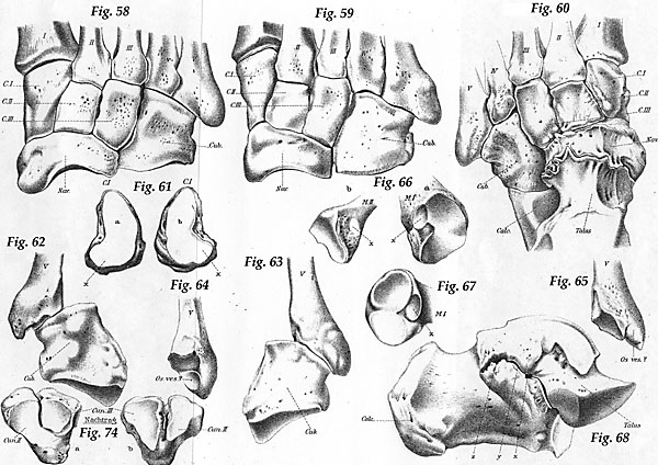
Figure 58:Lisfance'sche joint line. (tarsometatarsal joint). Dorsal view. Small bowing from the cuboid to cuneiform III.
Figure 59: Tarsometatarsal joint line. Dorsal view. Thinck bend crossing from the cuboid to metatarsal III.
Figure 60:Tarsometatarsal joint line. Dorsal view. Thick bend crossing from cuboid to cuneiform III. Coalescent talonavicular (pathologic?).
Figure 61: Special lower facet in the tarsometatarso I articulation. Distal joint surface of cuneiform I.
a: Rightb:Left
Figure 62:Os Vesalianum: the tubeosity of metatarsal V arises broodly over the cuboid.
Figure 63:Os Vesalianum: the tuberosity metatarsal V articulates in a special facet with the cuboid.
Figure 64:Os Vesalianum: the tuberosity metatarsal V articulates in a special facet with the cuboid.
Figure 65: Os Vesalianum set beyond a sharp furrow.
Figure 66:Articulation of intermetatarsum I/II.
a:Metatarsal I.b:Metatarsal II. x = facet of the intermetatarsal joint.
Figure 67: Articulation of intermetatarsum I/II. Proximal end of metatarsal I: the joint surface for cuneiform I is in a dorsal (d) and a plantar (pl) half is decayed; fibula next to the dorsal half of the facet. x - for the intermetatarsal joint.
Figure 68: Coalescence talocalcaneus. Talus and calcaneus in-situ, medial view. x = mouth of the anterior branch of the tarsal canal. y = accessory joint between talus and sustentaculum; z = coalescence off talus and posterior end of sustentaculum.
Figure 74: Supplement to tafel XIV. Synostosis between intercuneiform II/III.
a:Proximal.b: Distal view.
Section Top | Title Page
Please send us comments by filling out our Comment Form.
All contents copyright © 1995-2024 the Author(s) and Michael P. D'Alessandro, M.D. All rights reserved.
"Anatomy Atlases", the Anatomy Atlases logo, and "A digital library of anatomy information" are all Trademarks of Michael P. D'Alessandro, M.D.
Anatomy Atlases is funded in whole by Michael P. D'Alessandro, M.D. Advertising is not accepted.
Your personal information remains confidential and is not sold, leased, or given to any third party be they reliable or not.
The information contained in Anatomy Atlases is not a substitute for the medical care and advice of your physician. There may be variations in treatment that your physician may recommend based on individual facts and circumstances.
URL: http://www.anatomyatlases.org/