
Atlas of Human Anatomy in Cross Section: Section 1. Head and Neck
Ronald A. Bergman, Ph.D., Adel K. Afifi, M.D., Jean J. Jew, M.D., and Paul
C. Reimann, B.S.
Peer Review Status: Externally Peer Reviewed
|
Upper Left Quadrant |
Lower Left Quadrant |
Lower Right Quadrant |
Upper Right Quadrant |
|
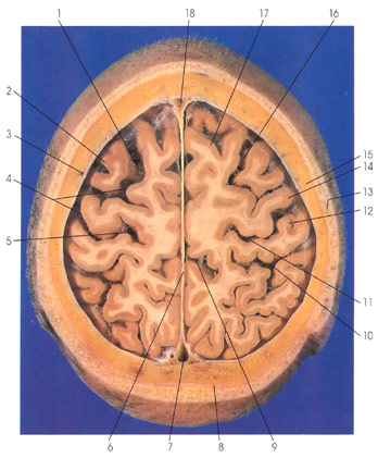
1. Superior frontal sulcus |
5. Central (rolandic) sulcus |
8. Galea aponeurotica |
12. Postcentral gyrus |
In this section (looking down), the superficial temporal artery (13) is seen within the scalp on the right side and the middle meningeal artery (3) is seen superficial to the dura mater on the left side. Skull fracture may injure the middle meningeal artery, leading to its rupture and a life threatening collection of arterial blood in the epidural space (epidural hemorrhage). The coronal suture (15) is seen between the frontal and parietal bones. The galea aponeurotica (8), an epicranial aponeurosis sandwiched between the connective tissue and loose areolar tissue layers of the scalp, is seen posteriorly. A dural fold, the falx cerebri (6), is seen in the interhemispheric fissure. Within the falx cerebri, rostrally and caudally, is the superior sagittal sinus (7, 18). In the frontal lobe the superior (17) and middle (2) frontal gyri are seen separated by the superior frontal sulcus (1, 16). The precentral gyrus (14) of the frontal lobe is separated from the postcentral gyrus (12) of the parietal lobe by the central (rolandic) sulcus (5, 11). The precentral gyrus comprises the primary motor cortex, whereas the postcentral gyrus comprises the primary somatosensory (somesthetic) cortex. Caudal to the postcentral gyrus is the postcentral sulcus (10). The marginal sulcus (9) is seen on the medial surface of the parietal lobe. Subarachnoid (cerebrospinal fluid-containing) spaces (4) are seen within sulci (1) and over the surface of each hemisphere.
Next Page | Previous Page | Section Top | Title Page
Please send us comments by filling out our Comment Form.
All contents copyright © 1995-2024 the Author(s) and Michael P. D'Alessandro, M.D. All rights reserved.
"Anatomy Atlases", the Anatomy Atlases logo, and "A digital library of anatomy information" are all Trademarks of Michael P. D'Alessandro, M.D.
Anatomy Atlases is funded in whole by Michael P. D'Alessandro, M.D. Advertising is not accepted.
Your personal information remains confidential and is not sold, leased, or given to any third party be they reliable or not.
The information contained in Anatomy Atlases is not a substitute for the medical care and advice of your physician. There may be variations in treatment that your physician may recommend based on individual facts and circumstances.
URL: http://www.anatomyatlases.org/