
Atlas of Human Anatomy in Cross Section: Section 1. Head and Neck
Ronald A. Bergman, Ph.D., Adel K. Afifi, M.D., Jean J. Jew, M.D., and Paul
C. Reimann, B.S.
Peer Review Status: Externally Peer Reviewed
|
Upper Left Quadrant |
Lower Left Quadrant |
Lower Right Quadrant |
Upper Right Quadrant |
|
1. Internal carotid a. |
8. Tentorium cerebelli |
13. Vermis of cerebellum |
20. Trigeminal nerve |
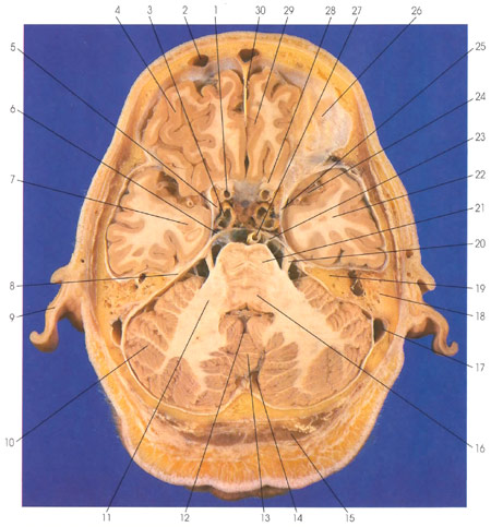
This is a section (looking down) through the frontal and temporal lobes, the pons, and the cerebellum. The frontal lobe is in the anterior cranial fossa, the temporal lobe is in the middle cranial fossa, and the cerebellum and pons are in the posterior cranial fossa. The falx cerebri (30) is seen in the interhemispheric fissure between the two frontal lobes (4). The orbital part of the frontal lobe (4, 26) and the gyrus rectus (29) are seen. The frontal sinus (2) is seen on the left side. Within the temporal lobe (22), the amygdaloid nucleus (7) is seen. Branches of the middle cerebral artery (24) are in the lateral (sylvian) fissure. Caudal to the frontal lobe (4) are the optic nerve (28) and the internal carotid artery (1, 3). The oculomotor nerve (5) is adjacent to the internal carotid artery (3). Caudal to the temporal lobe (22) is the petrous portion of the temporal bone (19) and the mastoid air cells (18). The sigmoid sinus (17) is seen within the dura. The tentorium cerebelli (8) is continuous with the posterior (6) and anterior (25) petroclinoid ligaments. In the posterior fossa, the cerebellum (10, 13) and pons (16, 21) are seen. The two are connected via the middle cerebellar peduncle (brachium pontis) (11). The trigeminal nerve (20) leaves the lateral surface of the pons, and the abducens nerve (23) leaves the ventral surface of the pons. The basilar artery (27) is ventral to the pons. The fourth ventricle (12) is between the pons and cerebellum. The cisterna magna (14) is caudal to the cerebellum. Outside the cranial cavity, the auricle (9) and the semispinalis capitis muscle (15) are seen.
Next Page | Previous Page | Section Top | Title Page
Please send us comments by filling out our Comment Form.
All contents copyright © 1995-2025 the Author(s) and Michael P. D'Alessandro, M.D. All rights reserved.
"Anatomy Atlases", the Anatomy Atlases logo, and "A digital library of anatomy information" are all Trademarks of Michael P. D'Alessandro, M.D.
Anatomy Atlases is funded in whole by Michael P. D'Alessandro, M.D. Advertising is not accepted.
Your personal information remains confidential and is not sold, leased, or given to any third party be they reliable or not.
The information contained in Anatomy Atlases is not a substitute for the medical care and advice of your physician. There may be variations in treatment that your physician may recommend based on individual facts and circumstances.
URL: http://www.anatomyatlases.org/