
Atlas of Human Anatomy in Cross Section: Section 1. Head and Neck
Ronald A. Bergman, Ph.D., Adel K. Afifi, M.D., Jean J. Jew, M.D., and Paul
C. Reimann, B.S.
Peer Review Status: Externally Peer Reviewed
|
Upper Left Quadrant |
Lower Left Quadrant |
Lower Right Quadrant |
Upper Right Quadrant |
|
1. Ethmoidal labyrinth |
9. Semicircular canals |
18. Choroid plexus in roof of fourth ventricle |
25. Pons |
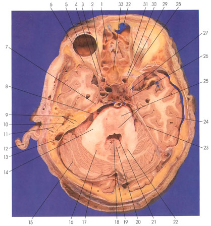
This is a section through the temporal lobe and brain stem. In the orbital cavity, the following structures are seen: lateral rectus muscle (6), eyeball (5), medial rectus muscle (2, 31), and superior rectus muscle (28). The optic nerve (30) is seen behind the orbit in close proximity to the internal carotid artery (29). In the midline rostrally are the frontal (32) and spheroid (33) sinuses. The ethmoidal labyrinth (1) is seen medial to the orbit. The oculomotor nerve (27) and the trigeminal (gasserian) ganglion (7) are seen medial to the temporal lobe (24). In the posterior fossa, the cerebellum (15, 20) and pons (25) are seen. The two are connected via the middle cerebellar peduncle (brachium pontis) (14). The fourth ventricle (21) is between the pons (25) and the vermis of the cerebellum (20).
The choroid plexus (18) is seen in the roof of the fourth ventricle (21). Deep within the cerebellum are the dentate nuclei (16, 22). The basilar artery (26) lies in a groove on the ventral surface of the pons (25). The trigeminal nerve (8) exits lateral to the pons (25). The tentorium cerebelli (23) is on each side of the cerebellum. The transverse (17) and sigmoid (13) sinuses and the confluence of sinuses (19) are seen. The anterior clinoid process (4) is seen lateral to the internal carotid artery (3). The mastoid air cells (11) are seen rostral to the sigmoid sinus (13) along with the middle ear (10) and the semicircular canals (9) of the inner ear. Part of the auricle (12) is seen on one side.
Next Page | Previous Page | Section Top | Title Page
Please send us comments by filling out our Comment Form.
All contents copyright © 1995-2024 the Author(s) and Michael P. D'Alessandro, M.D. All rights reserved.
"Anatomy Atlases", the Anatomy Atlases logo, and "A digital library of anatomy information" are all Trademarks of Michael P. D'Alessandro, M.D.
Anatomy Atlases is funded in whole by Michael P. D'Alessandro, M.D. Advertising is not accepted.
Your personal information remains confidential and is not sold, leased, or given to any third party be they reliable or not.
The information contained in Anatomy Atlases is not a substitute for the medical care and advice of your physician. There may be variations in treatment that your physician may recommend based on individual facts and circumstances.
URL: http://www.anatomyatlases.org/