
Atlas of Human Anatomy in Cross Section: Section 1. Head and Neck
Ronald A. Bergman, Ph.D., Adel K. Afifi, M.D., Jean J. Jew, M.D., and Paul
C. Reimann, B.S.
Peer Review Status: Externally Peer Reviewed
|
Upper Left Quadrant |
Lower Left Quadrant |
Lower Right Quadrant |
Upper Right Quadrant |
|
1. Nasal septum |
21. Spinal accessory (CN 11) nerve |
37. Occipital bone |
50. External auditory meatus |
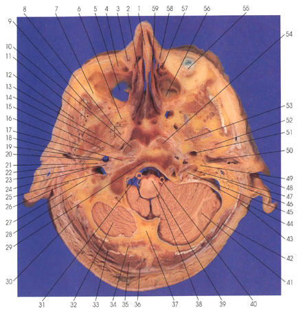
The following nerves can be identified: facial (CN 7) (27, 49), glossopharyngeal (CN 9), vagus (CN 10), spinal accessory (CN 11) (21, 23, 45), sympathetic trunk (20), and the infraorbital (7) nerve of the maxillary division of the trigeminal nerve (CN 5). This section cuts the medullary pyramids (39) and the cerebellar hemispheres (32, 41).
The muscles of mastication include the lateral pterygoid (13), masseter (12), temporalis (9), and the edge of the medial pterygoid (8). These four muscles are innervated by the trigeminal nerve (CN 5), mandibular division.
Vascular elements identified are internal carotid artery (19, 51), sigmoid sinus (44), vertebral arteries (31,40), internal jugular vein (25), superficial temporal artery (18), middle meningeal artery (15), deep temporal artery (11), and pterygoid plexus of veins (6).
The levator veli palatini muscle (14), which elevates the soft palate, is innervated by a pharyngeal branch of the vagus nerve. The tensor veli palatini (not seen in this section) is innervated by the motor part of the mandibular division of the trigeminal nerve (CN 5), which passes through the otic ganglion. This muscle is different from the other muscles of the palate, and this difference is explained by its development from the mandibular arch.
Muscles of the head and neck include the sternocleidomastoid (43) and trapezius (36), which are innervated by the spinal accessory nerve (CN 11); splenius capitis (30, 42) and semispinalis capitis (35), which are innervated by the lateral division of the posterior primary rami of upper cervical nerves; rectus capitis anterior (17), which is innervated by the ventral division of the first or first and second cervical nerves; and longus capitis (10), which is innervated by direct branches from the anterior division of cervical nerves. The suboccipital muscles seen here include rectus capitis posterior major (33) and minor (34). All four suboccipital muscles (two are not seen in this section) are innervated by the posterior primary ramus of the suboccipital (first cervical) nerve.
Next Page | Previous Page | Section Top | Title Page
Please send us comments by filling out our Comment Form.
All contents copyright © 1995-2024 the Author(s) and Michael P. D'Alessandro, M.D. All rights reserved.
"Anatomy Atlases", the Anatomy Atlases logo, and "A digital library of anatomy information" are all Trademarks of Michael P. D'Alessandro, M.D.
Anatomy Atlases is funded in whole by Michael P. D'Alessandro, M.D. Advertising is not accepted.
Your personal information remains confidential and is not sold, leased, or given to any third party be they reliable or not.
The information contained in Anatomy Atlases is not a substitute for the medical care and advice of your physician. There may be variations in treatment that your physician may recommend based on individual facts and circumstances.
URL: http://www.anatomyatlases.org/