
Atlas of Human Anatomy in Cross Section: Section 1. Head and Neck
Ronald A. Bergman, Ph.D., Adel K. Afifi, M.D., Jean J. Jew, M.D., and Paul
C. Reimann, B.S.
Peer Review Status: Externally Peer Reviewed
|
Upper Left Quadrant |
Lower Left Quadrant |
Lower Right Quadrant |
Upper Right Quadrant |
|
1. Clivus |
7. Mastoid air cells |
13. Anterior spinal a. |
19. Internal carotid a. |
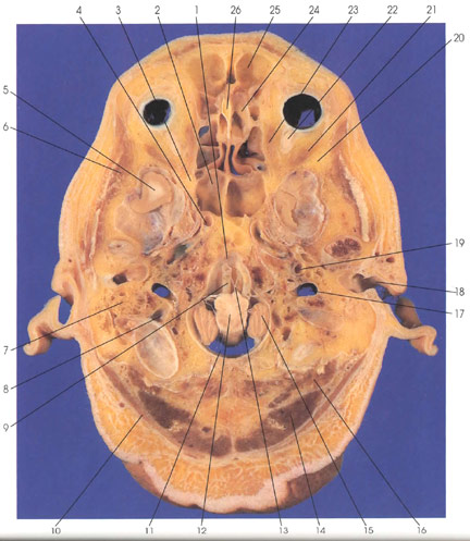
This is a section through the base of the cranial cavity. Rostrally in the midline is the crista galli (26) and lateral to it is the olfactory bulb (24). Rostral to the olfactory bulb (24) in the frontal bone is the frontal sinus (25). In the orbital cavity, the medial (23) and lateral (21) rectus muscles and the optic nerve (22) are seen. The optic canal (3) is caudal to the orbit. Within the middle cranial fossa is the temporal lobe (5) and anterolateral to it is the greater wing of the sphenoid bone (20). In the posterior cranial fossa, the medulla oblongata (12) and cerebellar tonsils (15) are seen. Ventral to the medulla is the vertebral artery (9) and its branch, the anterior spinal artery (13). The clivus (1) is also seen ventral to the medulla (12). Arachnoid trabeculae (11) are seen in the subarachnoid space around the medulla oblongata (12). Several major vessels are seen in this section: internal carotid artery (4, 19), vertebral artery (9), and jugular vein (17). The sigmoid venous sinus (8) and the sphenoid air sinus (2) are seen. Lateral to the sigmoid sinus (8) are the mastoid air cells (7). The external auditory meatus (18) is seen. Outside the cranial cavity, the following muscles are seen: rectus capitis posterior major (16), semispinalis capitis (14), splenius capitis (10), and temporalis (6).
Next Page | Previous Page | Section Top | Title Page
Please send us comments by filling out our Comment Form.
All contents copyright © 1995-2024 the Author(s) and Michael P. D'Alessandro, M.D. All rights reserved.
"Anatomy Atlases", the Anatomy Atlases logo, and "A digital library of anatomy information" are all Trademarks of Michael P. D'Alessandro, M.D.
Anatomy Atlases is funded in whole by Michael P. D'Alessandro, M.D. Advertising is not accepted.
Your personal information remains confidential and is not sold, leased, or given to any third party be they reliable or not.
The information contained in Anatomy Atlases is not a substitute for the medical care and advice of your physician. There may be variations in treatment that your physician may recommend based on individual facts and circumstances.
URL: http://www.anatomyatlases.org/