Anatomy Atlases: Atlas of Human Anatomy in Cross Section: Section 1. Head
and Neck
Atlas of Human Anatomy in Cross Section: Section 1. Head and Neck
Plate 1.24
Ronald A. Bergman, Ph.D., Adel K. Afifi, M.D., Jean J. Jew, M.D., and Paul
C. Reimann, B.S.
Peer Review Status: Externally Peer Reviewed
Upper Left Quadrant
Lower Left Quadrant
Lower Right Quadrant
Upper Right Quadrant
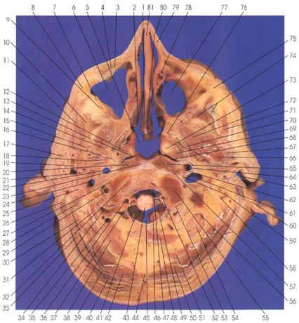
1. Nasal septum, vomer bone
2. Nasal cavity and mucous membrane
3. Infraorbital nerve
4. Zygomaticus major m.
5. Nasopharynx
6. Maxillary sinus
7. Maxilla
8. Zygomatic bone
9. Maxillary a.
10. Mandible
11. Masseter m.
12. Tensor veli palatini m.
13. Lingual nerve and superior pharyngeal constrictor m.
14. Trigeminal nerve (CN 7), mandibular division
15. Inferior alveolar a. and nerve
16. Auriculotemporal nerve and sphenomandibular ligament
17. Parotid gland
18. Middle meningeal a.
19. Facial nerve (CN 7)
20. Longus capitis m.
21. External carotid a.
22. Internal carotid a.
23. Anterior longitudinal ligament
24. Internal jugular v.
25. Facial nerve (CN 7)
26. Sternocleidomastoid m.
27. Occipital condyle
28. Digastric m., posterior belly
29. Vertebral a.
30. Longissimus capitis m.
31. Rectus capitis lateralis m.
32. First cervical vertebra, atlas
33. Longissimus capitis m.
34. Vertebral a.
35. Posterior vertebral venous plexus
36. Vertebral a.
37. Vertebral v.
38. Rectus capitis posterior major m.
39. Greater occipital nerve
40. Trapezius m.
41. Subarachnoid trabeculae
42. Semispinalis capitis m.
43. Medulla, lower (caudal) end
44. Ligamentum nuchae
45. Foramen magnum
46. Cerebellum
47. Tectorial membrane and cruciform ligament of atlas
48. Rectus capitis posterior minor m.
49. Posterior atlantooccipital membrane
50. Semispinalis capitis m.
51. Rectus capitis posterior major m.
52. Vertebral a.
53. First cervical vertebra, atlas
54. Cerebellum
55. Occipital condyle
56. Posterior cranial fossa
57. Tendon m. sternocleidomastoid and rectus capitis anterior m.
58. Mastoid air cells
59. Glossopharyngeal, vague, and spinal accessory nerves
60. Auricular elastic cartilage
61. Internal jugular v.
62. Cervical sympathetic trunk
63. External auditory meatus
64. Internal carotid a.
65. Sphenomandibular ligament and longus capitis m.
66. Mandible, condylar process
67. Temporomandibular ligament
68. Pharyngeal recess (of Rosenmueller) of nasopharynx
69. Levator veli palatini m.
70. Trigeminal nerve (CN 5), mandibular division
71. Masseter m.
72. Lateral pterygoid m.
73. Auditory (eustachian) tube
74. Temporalis tendon and m.
75. Zygomatic bone
76. Medial pterygoid m.
77. Maxillary sinus
78. Nasolacrimal duct
79. Lateral nasal cartilages
80. Mucous membrane, nasal septum, and lateral wall
81. Septal nasal cartilage
This section passes through the maxillary sinus (6, 77), nasopharynx (5), nasal
septum (80), zygomatic bone (8, 75), medial (76) and lateral (72) pterygoid muscles,
masseter (11, 71), condylar process of the mandible (66), occipital condyles (55),
foramen magnum (45), inferior end of the left posterior cranial fossa (56) and
cerebellum (54), and the parotid gland (17).
The vertebral artery (34, 36) is seen crossing transversely from the foremen
of the transverse process to enter the foremen magnum of the right side. The
artery has already entered the foremen magnum on the left side (52).
The sphenomandibular ligament (16) is a relatively thin, loose band of connective
tissue. It is attached superiorly to the spine of the sphenoid and the region
of the petrotympanic fissure. Inferiorly, it is attached to the lingula of the
mandible and the superior end of the mylohyoid groove, where it is pierced by
the mylohyoid nerve. The sphenomandibular ligament is separated from the temporomandibular
joint and ramus of the mandible by the maxillary artery and vein, the inferior
alveolar nerve (15) and artery, auriculotemporal nerve (16), and the middle
meningeal artery (18).
The sphenomandibular ligament (16) is a fibrous remnant derived, in part, from
Meckel's cartilage.
"Anatomy Atlases", the Anatomy Atlases logo, and "A digital library of anatomy information" are all Trademarks of Michael P. D'Alessandro, M.D.
Anatomy Atlases is funded in whole by Michael P. D'Alessandro, M.D. Advertising is not accepted.
Your personal information remains confidential and is not sold, leased, or given to any third party be they reliable or not.
The information contained in Anatomy Atlases is not a substitute for the medical care and advice of your physician. There may be variations in treatment that your physician may recommend based on individual facts and circumstances.