
Atlas of Human Anatomy in Cross Section: Section 1. Head and Neck
Ronald A. Bergman, Ph.D., Adel K. Afifi, M.D., Jean J. Jew, M.D., and Paul
C. Reimann, B.S.
Peer Review Status: Externally Peer Reviewed
|
Upper Left Quadrant |
Lower Left Quadrant |
Lower Right Quadrant |
Upper Right Quadrant |
|
1. Lingual septum |
7. Tongue, root |
27. Vertebra, third cervical lamina |
51. Masseter m. |
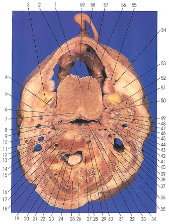
This section passes through the inferior edge of the nose (58), the tongue (57), mandible (3, 49), oral pharynx (32), first cervical disk (23), third cervical vertebral body (19), lamina (27), and transverse process (16).
The section cuts the inferior alveolar (CN 5) (52), lingual (CN 5) (50), greater auricular (44), facial (CN 7) (43), glossopharyngeal (CN 9) (41), hypoglossal (CN 12) (38), superior laryngeal (CN 10) (36), vagus (CN 10) (35), and third cervical (12) nerves.
The transverse linguae muscle (57) is well seen in this section. Also note the cervical enlargement of the spinal cord (24).
Two of the three pharyngeal constrictor muscles can be recognized in this section: rhe superior constrictor muscle (33) and the edge of the overlapping middle constrictor muscle (14). The middle constrictor arises, in part, from the stylohyoid ligament (8,40) and inserts into the connective tissue pharyngeal raphe (32) in the middle of the posterior wall of the pharynx.
The first cervical intervertebral disk (23) occurs between the axis and the third cervical vertebra. No disk is found between the atlas and axis.
Next Page | Previous Page | Section Top | Title Page
Please send us comments by filling out our Comment Form.
All contents copyright © 1995-2025 the Author(s) and Michael P. D'Alessandro, M.D. All rights reserved.
"Anatomy Atlases", the Anatomy Atlases logo, and "A digital library of anatomy information" are all Trademarks of Michael P. D'Alessandro, M.D.
Anatomy Atlases is funded in whole by Michael P. D'Alessandro, M.D. Advertising is not accepted.
Your personal information remains confidential and is not sold, leased, or given to any third party be they reliable or not.
The information contained in Anatomy Atlases is not a substitute for the medical care and advice of your physician. There may be variations in treatment that your physician may recommend based on individual facts and circumstances.
URL: http://www.anatomyatlases.org/