Anatomy Atlases(tm) : A digital library of anatomy information

Home | About | FAQ | Reviews | Search

Anatomy Atlases: Atlas of Human Anatomy in Cross Section: Section 2. Neck, Shoulders, Upper Arm, and Upper Thorax (Lungs)

Atlas of Human Anatomy in Cross Section: Section 2. Neck, Shoulders, Upper Arm, and Upper Thorax (Lungs)

Plate 2.6

Ronald A. Bergman, Ph.D., Adel K. Afifi, M.D., Jean J. Jew, M.D., and Paul C. Reimann, B.S.
Peer Review Status: Externally Peer Reviewed


Plate 2.6

Upper Left Quadrant

Lower Left Quadrant

Lower Right Quadrant

Upper Right Quadrant

1. Recurrent laryngeal nerve
2. Anterior jugular v.
3. Tendon m. sternocleidomastoid
4. Parathyroid gland
5. Inferior thyroid a. and v.
6. Internal jugular v. and common carotid a.
7. Phrenic nerve
8. Subclavian a.
9. Clavicle
10. Brachial plexus
11. Anterior scalene m.
12. Subclavius m.
13. Pectoralis major m
14. Brachial plexus
15. Axillary a.
16. Brachial plexus, C8 nerve root
17. Cephalic v. and rib 1
18. Brachial plexus of nerves
19. Pectoralis major m.
20. Tendon m. biceps brachii, long head
21. Axillary fat
22. Humerus
23. Tendon m. Iatissimus dorsi
24. Lung, right upper lobe, apical segment, apex
25. Tendon m. teres major

26. Scapula
27. Posterior humeral circumflex a. and v.
28. Rib 2
29. Subscapular nerve
30. Parietal pleura
31. Teres minor m.
32. Suprascapular a. and v., infraspinatus brs.
33. Scapula
34. Subscapularis m.
35. Rib 3
36. Scapula
37. Thoracolumbar fascia
38. Serratus anterior m.
39. Infraspinatus m.
40. Transverse cervical a. and v.
41. Costovertebral articulation, T3
42. Scapula, medial border
43. Rhomboideus minor m.
44. Rhomboideus major m.
45. Semispinalis capitis m.
46. Trapezius m.
47. Vertebra, T2 body
48. Basivertebral v.

49. Supraspinous ligament
50. Vertebra, T1 spinous process
51. Thoracic spinal cord
52. Trapezius m.
53. Rhomboideus major m.
54. Semispinalis capitis m.
55. Multifidus m.
56. Rhomboideus minor m.
57. Scapula, medial border
58. Dorsal scapular nerve (nerve to rhomboids)
59. Subarachnoid space
60. Intervertebral v., anterior transverse br.
61. Subscapularis m.
62. Intercostal mm.
63. Infraspinatus m.
64. Serratus anterior m.
65. Teres minor m.
66. Rib 3
67. Scapula
68. Axillary nerve
69. Triceps brachii, long head
70. Scapula
71. Posterior humeral circumflex a., brs.
72. Intercostal mm.
73. Axillary nerve (splayed)
74. Deltoid m.

75. Subscapularis m.
76. Rib 2
77. Humerus, lesser tubercle
78. Humerus, greater tubercle
79. Tendon m. biceps brachii, long head in intertubercular groove of humerus
80. Lateral pectoral nerve entering pectoralis major m.
81. Lung, left upper lobe, upper division, apical posterior segment
82. Coracobrachialis and biceps brachii, short head mm.
83. Parietal pleura, pleural dome
84. Pectoral is minor m.
85. Cephalic v.
86. Brachial plexus of nerves
87. Rib 1
88. Brachial plexus of nerves
89. Internal jugular v.
90. Brachial plexus of nerves, dorsal root ganglion
91. Subclavius m.
92. External jugular v.
93. Brachial plexus of nerves
94. Subclavian a.
95. Clavicle
96. Anterior scalene m.
97. Phrenic nerve
98. Vagus nerve, CN 10
99. Thoracic duct and intervertebral disk, T1
100. Internal jugular v.
101. Common carotid a.
102. Recurrent laryngeal nerve and vertebra, T1
103. Sternohyoid m.
104. Thyroid gland
105. Anterior jugular v.
106. Esophagus
107. Trachea

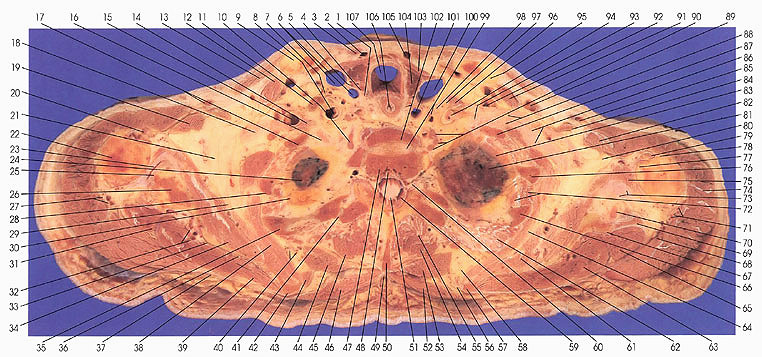
This section passes through the thyroid gland (104), esophagus (106) (first appearance), first (102) and second (47) thoracic vertebrae, brachial plexus of nerves (10,16,86,88,90,93), scapula (26,33,36,42,57,67), clavicle (9,95), and the pleural dome and lungs (24,81).

In this section, the transition of pharynx to esophagus occurs.

Important blood vessels in the neck include the common carotid (6,101) and subclavian arteries (8,94), the anterior (2,105) and internal (6,100) jugular veins, and the transverse cervical (40), suprascapular (32), and inferior thyroid (5) arteries and veins.

The cranial end of the thorax is seen in this section. The uppermost part of the lungs can be seen and the following bronchopulmonary segments can be identified: the left lung, upper lobe, upper division, apical posterior segment (81), and the right lung, upper lobe, apical segment (24).

The serratus anterior muscle (38,64) usually arises from the first through eighth or ninth ribs and inserts onto the costal surface of the scapula, from its superior to its inferior angle.

The brachial plexus (86,88,90,93) of nerves is well seen on the left side where the roots of cervical nerves 5 through 8 and the first thoracic nerve have passed behind the anterior scalene muscle (96). The subclavian artery (94) takes the same route, behind the anterior scalene muscle and over the first rib, after which it is renamed the axillary artery. The vessel makes this transition in this section.

The humerus is cut just below its articulation with the scapula; its greater (78) and lesser (77) tubercles are seen. The two tubercles are separated by the intertubercular groove housing the tendon of the long head of biceps brachii (79).

Next Page | Previous Page | Section Top | Title Page


Home | About Us | FAQ | Reviews | Contact Us | Search

Anatomy Atlases is curated by Michael P. D'Alessandro, M.D. and Ronald A. Bergman, Ph.D.

Please send us comments by filling out our Comment Form.

All contents copyright © 1995-2024 the Author(s) and Michael P. D'Alessandro, M.D. All rights reserved.

"Anatomy Atlases", the Anatomy Atlases logo, and "A digital library of anatomy information" are all Trademarks of Michael P. D'Alessandro, M.D.

Anatomy Atlases is funded in whole by Michael P. D'Alessandro, M.D. Advertising is not accepted.

Your personal information remains confidential and is not sold, leased, or given to any third party be they reliable or not.

The information contained in Anatomy Atlases is not a substitute for the medical care and advice of your physician. There may be variations in treatment that your physician may recommend based on individual facts and circumstances.

URL: http://www.anatomyatlases.org/