
Atlas of Human Anatomy in Cross Section: Section 3. Middle Thorax (Heart and Lungs)
Ronald A. Bergman, Ph.D., Adel K. Afifi, M.D., Jean J. Jew, M.D., and Paul
C. Reimann, B.S.
Peer Review Status: Externally Peer Reviewed
|
Upper Left Quadrant |
Lower Left Quadrant |
Lower Right Quadrant |
Upper Right Quadrant |
|
1. Rib 3 |
12. Lung, right upper lobe, posterior segment |
31. Interspinous ligament |
45. Intercostal mm. and left interlobar (oblique) fissure |
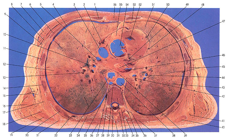
This section passes through the lower third of the seventh thoracic vertebra (32), the zygapophyseal joint (33) with its superior and inferior articular processes, the interspinous ligament (31), and the body of the sternum (56) anteriorly. It cuts ribs 7 (20), 6 (14), 5 (11), 4 (6), and 3 (1). On the left side, the right coronary artery (55), ascending aorta (54), right ventricle (52), left anterior descending coronary artery and vein (50), left coronary artery (49), pericardiacophrenic artery and phrenic nerve (48), and left atrium (47) are seen. The thoracic aorta (38) and hemiazygos vein (35) are also seen. The section cuts the left lung, upper lobe, upper division, anterior (51); the left upper lobe, upper division, apical posterior (46); and the left lower lobe, superior (43) bronchopulmonary segments.
On the right side, the superior vena cave (3), right pulmonary artery (5), lateral thoracic artery and vein (9), thoracodorsal artery and vein (15), primary division of the right bronchus (10), and the esophagus (23) can be identified. The following right bronchopulmonary segments have been identified: right lower lobe, superior (23); right upper lobe, posterior (12); and right upper lobe, anterior (3).
A dorsal root ganglion in the intervertebral foramen (28), an intercostal neurovascular bundle (22), and the nerve supply to the serratus anterior muscle (17), the long thoracic nerve (15), is seen.
The teres major muscle (18, 41) makes its last appearance in this section.
Next Page | Previous Page | Section Top | Title Page
Please send us comments by filling out our Comment Form.
All contents copyright © 1995-2024 the Author(s) and Michael P. D'Alessandro, M.D. All rights reserved.
"Anatomy Atlases", the Anatomy Atlases logo, and "A digital library of anatomy information" are all Trademarks of Michael P. D'Alessandro, M.D.
Anatomy Atlases is funded in whole by Michael P. D'Alessandro, M.D. Advertising is not accepted.
Your personal information remains confidential and is not sold, leased, or given to any third party be they reliable or not.
The information contained in Anatomy Atlases is not a substitute for the medical care and advice of your physician. There may be variations in treatment that your physician may recommend based on individual facts and circumstances.
URL: http://www.anatomyatlases.org/