
Atlas of Human Anatomy in Cross Section: Section 4. Upper Limb
Ronald A. Bergman, Ph.D., Adel K. Afifi, M.D., Jean J. Jew, M.D., and Paul
C. Reimann, B.S.
Peer Review Status: Externally Peer Reviewed
|
Upper Left Quadrant |
Lower Left Quadrant |
Lower Right Quadrant |
Upper Right Quadrant |
|
1. Flexor carpi radialis m. |
8. Extensor carpi radialis brevis m. |
16. Abductor pollicis longus m. |
22. Flexor carpi ulnaris m. |
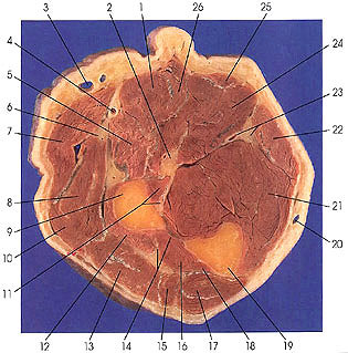
This section passes through the upper third of the forearm distal to the insertion of the arm muscles. The medially placed basilic (20) and the laterally placed cephalic (3) veins are seen.
The belly of palmaris longus (25) is decreasing in size and is tendinous in the next section.
Pronator teres muscle (5) arises from two heads. The humeral or chief head originates from the superior half of the ventral surface of the medial epicondyle and from the overlying fascia and intermuscular septa between it and the medial head of the triceps and flexor carpi radialis (26). The ulnar or deep head (accessory head) arises by an aponeurotic band from the medial border of the coronoid process of the ulna, medial to the tendon of the brachialis muscle. It is important to note that a fibrous arch extends between the humeral and ulnar heads through which the median nerve passes. At this point the median nerve can become trapped and compromised. The muscle inserts by a broad tendon that winds around the ventral surface of the radius (9) and attaches onto the middle third of its lateral surface. The muscle is innervated by a branch from the median nerve (2) before it passes between the two heads of the muscle. The nerve enters the proximal part of the middle third of the main belly of the muscle on its deep surface near the radial border. The branch to the ulnar head is proximal to its fusion with the humeral head.
Next Page | Previous Page | Section Top | Title Page
Please send us comments by filling out our Comment Form.
All contents copyright © 1995-2024 the Author(s) and Michael P. D'Alessandro, M.D. All rights reserved.
"Anatomy Atlases", the Anatomy Atlases logo, and "A digital library of anatomy information" are all Trademarks of Michael P. D'Alessandro, M.D.
Anatomy Atlases is funded in whole by Michael P. D'Alessandro, M.D. Advertising is not accepted.
Your personal information remains confidential and is not sold, leased, or given to any third party be they reliable or not.
The information contained in Anatomy Atlases is not a substitute for the medical care and advice of your physician. There may be variations in treatment that your physician may recommend based on individual facts and circumstances.
URL: http://www.anatomyatlases.org/