
Atlas of Human Anatomy in Cross Section: Section 4. Upper Limb
Ronald A. Bergman, Ph.D., Adel K. Afifi, M.D., Jean J. Jew, M.D., and Paul
C. Reimann, B.S.
Peer Review Status: Externally Peer Reviewed
|
Upper Left Quadrant |
Lower Left Quadrant |
Lower Right Quadrant |
Upper Right Quadrant |
|
1. First lumbrical m. |
8. Tendon m. extensor pollicis longus |
15. Dorsal interosseous m. |
22. Palmar metacarpal a. |
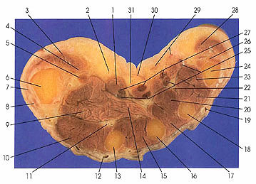
This section passes through the distal ends of the thenar (3, 4) and hypothenar (26, 28) muscles and the first metacarpal bone (6). Note the four lumbrical muscles (25). Palmar metacarpal arteries (11, 22) and superficial palmer branches arise from the radial artery.
The palmer metacarpal arteries are three in number, arise from the convexity of the deep arch, and extend distally in the second, third, and fourth interosseous spaces on the interosseous muscles deep to the palmer interosseous fascia They end by uniting with the palmer common digital arteries from the superficial (ulnar) arch. These arteries supply the interosseous muscles and bones and the second, third, and fourth lumbrical muscles. The superficial palmer branch of the radial arises from the distal end of the radial artery at the level of the styloid process of the radius. It perforates the deep layer of the palmer carpal ligament and enters the thenar compartment, passing across the thenar muscles and supplying them. It may join with the superficial branch of the ulnar artery to complete the superficial palmer arch in about one third of cases. It may also terminate in the thenar muscles or continue into the palm, supplying the radial digits without uniting with the superficial palmer arch.
Next Page | Previous Page | Section Top | Title Page
Please send us comments by filling out our Comment Form.
All contents copyright © 1995-2024 the Author(s) and Michael P. D'Alessandro, M.D. All rights reserved.
"Anatomy Atlases", the Anatomy Atlases logo, and "A digital library of anatomy information" are all Trademarks of Michael P. D'Alessandro, M.D.
Anatomy Atlases is funded in whole by Michael P. D'Alessandro, M.D. Advertising is not accepted.
Your personal information remains confidential and is not sold, leased, or given to any third party be they reliable or not.
The information contained in Anatomy Atlases is not a substitute for the medical care and advice of your physician. There may be variations in treatment that your physician may recommend based on individual facts and circumstances.
URL: http://www.anatomyatlases.org/