
Atlas of Human Anatomy in Cross Section: Section 6. Pelvis, Perineum, Hip, and Upper Thigh: Male
Ronald A. Bergman, Ph.D., Adel K. Afifi, M.D., Jean J. Jew, M.D., and Paul
C. Reimann, B.S.
Peer Review Status: Externally Peer Reviewed
|
Upper Left Quadrant |
Lower Left Quadrant |
Lower Right Quadrant |
Upper Right Quadrant |
|
1. Transverse colon |
8. Genitofemoral nerve |
25. Nerve roots and filum terminale; thecal sac |
40. Iliohypogastric nerve |
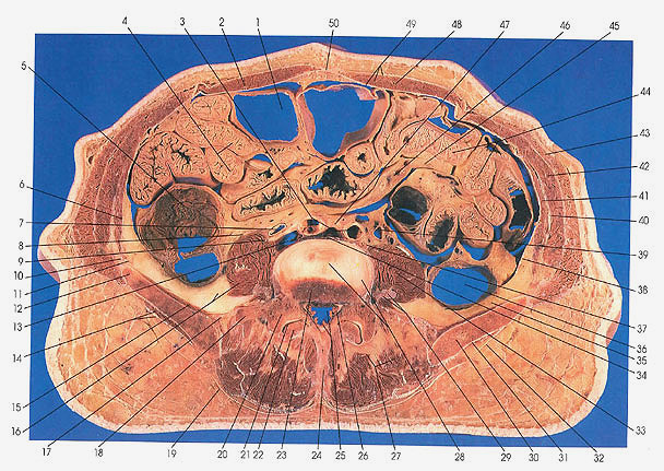
This section passes through the upper part of the fifth lumbar vertebra (23) and the lower part of the L4/L5 intervertebral disk (28).
At this level the bifurcation of the aorta has taken place, giving rise to the common iliac arteries (3). The inferior vena cave (13) is seen for the last time.
Gluteus medius (14, 31) and iliacus (15) muscles are seen for the first time.
The intestinal coils of the small intestine are cut many times: on the left side the jejunum (44) and on the right side the ileum (4). The retroperitoneal descending colon lying on the posterior abdominal wall is cut transversely (36). The ascending colon (5) is also seen. Although there is disagreement about where the sigmoid colon begins, Anson places it at the level of the superior aperture of the lesser or true pelvis (see Plate 6.5). Other authors (e.g., Poirier and Charpy, Spalteholtz, and Eycleshymer and Schoemaker) consider the beginning of the sigmoid to be below the place where the colon is in front of the iliac vessels and the left vas deferens. Still others consider the point of demarcation to be the crest of the ilium.
Next Page | Previous Page | Section Top | Title Page
Please send us comments by filling out our Comment Form.
All contents copyright © 1995-2024 the Author(s) and Michael P. D'Alessandro, M.D. All rights reserved.
"Anatomy Atlases", the Anatomy Atlases logo, and "A digital library of anatomy information" are all Trademarks of Michael P. D'Alessandro, M.D.
Anatomy Atlases is funded in whole by Michael P. D'Alessandro, M.D. Advertising is not accepted.
Your personal information remains confidential and is not sold, leased, or given to any third party be they reliable or not.
The information contained in Anatomy Atlases is not a substitute for the medical care and advice of your physician. There may be variations in treatment that your physician may recommend based on individual facts and circumstances.
URL: http://www.anatomyatlases.org/