
Atlas of Human Anatomy in Cross Section: Section 6. Pelvis, Perineum, Hip, and Upper Thigh
Ronald A. Bergman, Ph.D., Adel K. Afifi, M.D., Jean J. Jew, M.D., and Paul
C. Reimann, B.S.
Peer Review Status: Externally Peer Reviewed
|
Upper Left Quadrant |
Lower Left Quadrant |
Lower Right Quadrant |
Upper Right Quadrant |
|
1. Ileum |
12. Gluteus medius m. |
23. Pudendal nerve and sigmoid colon |
32. Gluteus medius m. |
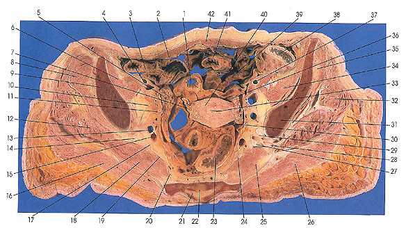
This section passes through the rectus abdominis muscles (42), suspensory ligament of the right ovary (5), uterus (40), ilium (9, 31), and sacrum (22).
On the left side, the peroneal (29) and tibial (27) divisions of the sciatic nerve are seen separated by the superior gluteal artery and vein in the sciatic foremen (28).
A portion of the broad ligament (30, 36) is seen covering the uterus (40). The uterine artery is seen on the right lateral surface of the uterus. The uterine cavity (33) is seen as a narrow slit in the thick body of the organ.
The pudendal nerves (20, 23) are seen near their origin from sacral nerves 2, 3, and 4. The pudendal nerve is variable and may arise from nerves S1 through S5, but usually from three nerves.
Next Page | Previous Page | Section Top | Title Page
Please send us comments by filling out our Comment Form.
All contents copyright © 1995-2024 the Author(s) and Michael P. D'Alessandro, M.D. All rights reserved.
"Anatomy Atlases", the Anatomy Atlases logo, and "A digital library of anatomy information" are all Trademarks of Michael P. D'Alessandro, M.D.
Anatomy Atlases is funded in whole by Michael P. D'Alessandro, M.D. Advertising is not accepted.
Your personal information remains confidential and is not sold, leased, or given to any third party be they reliable or not.
The information contained in Anatomy Atlases is not a substitute for the medical care and advice of your physician. There may be variations in treatment that your physician may recommend based on individual facts and circumstances.
URL: http://www.anatomyatlases.org/