
Atlas of Human Anatomy in Cross Section: Section 6. Pelvis, Perineum, Hip, and Upper Thigh
Ronald A. Bergman, Ph.D., Adel K. Afifi, M.D., Jean J. Jew, M.D., and Paul
C. Reimann, B.S.
Peer Review Status: Externally Peer Reviewed
|
Upper Left Quadrant |
Lower Left Quadrant |
Lower Right Quadrant |
Upper Right Quadrant |
|
1. Urinary bladder |
18. Acetabulum |
31. Rectouterine pouch (of Douglas) |
40. Pericervical fascia |
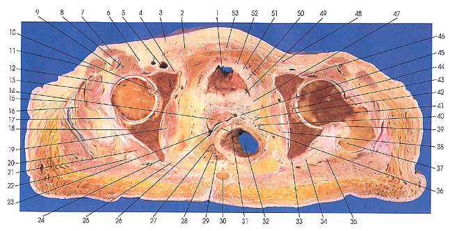
This section passes through the tendon of rectus abdominis (53), urinary bladder (1), the heads of the femora (43, 44), left greater trochanter of the femur (37), anterior lip of the uterus (14), vagina (23), ischial spines (24, 33), ampulla of the rectum (32), and the coccyx (30).
Various parts of the supporting fascia and ligaments of the uterus are identified in this section. They lie below the broad ligament identified earlier and are as follows: pubovesical fascia (52), perivesical fascia (50), uterovesical ligament (47), cardinal ligament (cervical ligament of uterus) (42), pericervical fascia (40), and uterorectal ligament (29).
The cardinal ligament is not a discrete structure but rather tough fibrous connective tissue that surrounds pelvic blood vessels and especially the uterine artery as it approaches the uterus.
Next Page | Previous Page | Section Top | Title Page
Please send us comments by filling out our Comment Form.
All contents copyright © 1995-2024 the Author(s) and Michael P. D'Alessandro, M.D. All rights reserved.
"Anatomy Atlases", the Anatomy Atlases logo, and "A digital library of anatomy information" are all Trademarks of Michael P. D'Alessandro, M.D.
Anatomy Atlases is funded in whole by Michael P. D'Alessandro, M.D. Advertising is not accepted.
Your personal information remains confidential and is not sold, leased, or given to any third party be they reliable or not.
The information contained in Anatomy Atlases is not a substitute for the medical care and advice of your physician. There may be variations in treatment that your physician may recommend based on individual facts and circumstances.
URL: http://www.anatomyatlases.org/