
Atlas of Human Anatomy in Cross Section: Section 6. Pelvis, Perineum, Hip, and Upper Thigh
Ronald A. Bergman, Ph.D., Adel K. Afifi, M.D., Jean J. Jew, M.D., and Paul
C. Reimann, B.S.
Peer Review Status: Externally Peer Reviewed
|
Upper Left Quadrant |
Lower Left Quadrant |
Lower Right Quadrant |
Upper Right Quadrant |
|
1. Ligamentum teres (round ligament) of uterus |
14. Obturator externus m. |
24. Coccyx (last appearance) |
35. Gluteus medius m. |
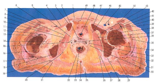
This section passes through the superior aspect of the pubic bones (3, 47), the lower portion of the left acetabulum at the insertion of the ligamentum teres femoris (40). Portions of the greater (32) and lesser (16) trochanters are seen. The ischial tuberosity (18) is seen on the right side. The anus (23) with its smooth muscle internal anal sphincter (22), and elements of levator ani muscle (30) can be identified.
The vagina (36) is located between the urinary bladder (48) and the anus (23).
The coccyx (24) makes its last appearance in this section.
The obturator membrane (7), closing the obturator foremen, is covered by the obturator internus muscle (10) and is seen on the right side.
Note the highly vascular vesical plexus of veins (2) (between the bladder [48] and vagina [36]) and the uterovaginal plexus of veins (33).
Next Page | Previous Page | Section Top | Title Page
Please send us comments by filling out our Comment Form.
All contents copyright © 1995-2024 the Author(s) and Michael P. D'Alessandro, M.D. All rights reserved.
"Anatomy Atlases", the Anatomy Atlases logo, and "A digital library of anatomy information" are all Trademarks of Michael P. D'Alessandro, M.D.
Anatomy Atlases is funded in whole by Michael P. D'Alessandro, M.D. Advertising is not accepted.
Your personal information remains confidential and is not sold, leased, or given to any third party be they reliable or not.
The information contained in Anatomy Atlases is not a substitute for the medical care and advice of your physician. There may be variations in treatment that your physician may recommend based on individual facts and circumstances.
URL: http://www.anatomyatlases.org/