
Atlas of Human Anatomy in Cross Section: Section 7. Lower Limb
Ronald A. Bergman, Ph.D., Adel K. Afifi, M.D., Jean J. Jew, M.D., and Paul
C. Reimann, B.S.
Peer Review Status: Externally Peer Reviewed
|
Upper Left Quadrant |
Lower Left Quadrant |
Lower Right Quadrant |
Upper Right Quadrant |
|
1. Profundus femoris a. and v. |
8. Gracilis m. |
15. Semitendinosus m. |
20. Vastus lateralis m. |
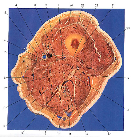
The muscles of the thigh are arranged in three groups based on their position and nerve supply: (a) The muscles on the front of the thigh (anterior or extensor group) are supplied by the femoral nerve (3). These muscles are the quadriceps femoris (rectus femoris) (24) and the three vasti (2, 20, 23) and sartorius (5). The second group of muscles are on the medial side of the thigh (adductor group) and are supplied by the obturator nerve (9). These include pectineus (see Section 6), gracilis (8), adductor longus (see Section 6), adductor brevis (6), adductor magnus (11), and obturator externus (see Section 6). (c) The third group of muscles are on the back of the thigh (posterior, flexor, or hamstring group) and are supplied by the sciatic nerve (18). These muscles are the biceps femoris (16, 17), semitendinosus (15), and semimembranosus (12).
The linea aspera of the femur (22) provides attachment for the vastus medialis (2), adductor longus (see Section 6), adductor magnus (11), adductor brevis (6), short head of biceps femoris (17), and vastus lateralis (20) muscles.
The lateral intermuscular septum (19) separating the anterior extensor group of muscles from the posterior flexor group and the important fascia lata (10) are seen.
The great saphenous vein (7), femoral artery and vein (4), perforating artery and vein (3), and the deep femoral artery and vein (1) are identified.
Next Page | Section Top | Title Page
Please send us comments by filling out our Comment Form.
All contents copyright © 1995-2024 the Author(s) and Michael P. D'Alessandro, M.D. All rights reserved.
"Anatomy Atlases", the Anatomy Atlases logo, and "A digital library of anatomy information" are all Trademarks of Michael P. D'Alessandro, M.D.
Anatomy Atlases is funded in whole by Michael P. D'Alessandro, M.D. Advertising is not accepted.
Your personal information remains confidential and is not sold, leased, or given to any third party be they reliable or not.
The information contained in Anatomy Atlases is not a substitute for the medical care and advice of your physician. There may be variations in treatment that your physician may recommend based on individual facts and circumstances.
URL: http://www.anatomyatlases.org/