
Atlas of Human Anatomy in Cross Section: Section 7. Lower Limb
Ronald A. Bergman, Ph.D., Adel K. Afifi, M.D., Jean J. Jew, M.D., and Paul
C. Reimann, B.S.
Peer Review Status: Externally Peer Reviewed
|
Upper Left Quadrant |
Lower Left Quadrant |
Lower Right Quadrant |
Upper Right Quadrant |
|
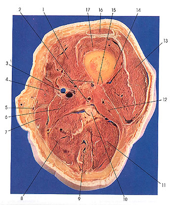
1. Vastus medialis m. |
5. Great saphenous v. |
10. Sciatic nerve |
13. Vastus lateralis m. |
This section is three sections (3 cm) below the preceding one. Rectus femoris (16) is decreasing in size and makes its last appearance in this section. It becomes tendinous, and this will be seen in the next cut. The tendon of rectus femoris is named the quadriceps tendon. The sciatic nerve (10) is also seen for the last time in this section. It will divide into its component tibial and common peroneal nerves.
The body or shaft of the femur is almost cylindric but it is slightly flattened anteriorly and strengthened posteriorly by a longitudinal ridge, the linea aspera (15). The linea aspera (rough line) extends along the middle third of the body of the femur and presents a medial (2) and lateral (15) lip separated by a narrow (plateau) interval. When followed onto the proximal third of the body, the lips diverge. The lateral lip becomes continuous with the gluteal tuberosity and ends at the base of the greater trochanter. The gluteal tuberosity, when prominently developed, is known as the third trochanter. The medial lip curves distal to the lesser trochanter, where it becomes continuous with the intertrochanteric line. Toward the distal third of the body of the femur, the medial and lateral lips of the linea aspera again diverge and extend to the condyles, enclosing between them the triangular surface of bone, the popliteal surface (planum popliteum), which forms the proximal part of the floor of the popliteal fossa. The lateral lip is more prominent and terminates distally in the lateral epicondyle. The medial lip, interrupted where the femoral vessels contact bone, terminates in the adductor tubercle, a sharp projection at the summit of the medial epicondyle, affording an insertion for the adductor magnus muscle.
From the medial lip of linea aspera and the intertrochanteric line, vastus medialis (1) arises and, from the lateral lip and lateral to the gluteal tuberosity, vastus lateralis (13) arises. The insertion of adductor magnus muscle (6) extends from the gluteal tuberosity through the length of the linea aspera, between its lips. Between adductor magnus (6) and vastus medialis (1), four muscles are inserted: proximally, pectineus (ee Section 6) and iliacus (see Section 6); next, adductor brevis (Sections 6 and 7); and distally, adductor longus (see Sections 6 and 7).
The short head of biceps femoris (12) takes its origin from the distal two-thirds of the lateral lip of linea aspera and the supracondylar ridge.
Next Page | Previous Page | Section Top | Title Page
Please send us comments by filling out our Comment Form.
All contents copyright © 1995-2024 the Author(s) and Michael P. D'Alessandro, M.D. All rights reserved.
"Anatomy Atlases", the Anatomy Atlases logo, and "A digital library of anatomy information" are all Trademarks of Michael P. D'Alessandro, M.D.
Anatomy Atlases is funded in whole by Michael P. D'Alessandro, M.D. Advertising is not accepted.
Your personal information remains confidential and is not sold, leased, or given to any third party be they reliable or not.
The information contained in Anatomy Atlases is not a substitute for the medical care and advice of your physician. There may be variations in treatment that your physician may recommend based on individual facts and circumstances.
URL: http://www.anatomyatlases.org/