
Atlas of Human Anatomy in Cross Section: Section 7. Lower Limb
Ronald A. Bergman, Ph.D., Adel K. Afifi, M.D., Jean J. Jew, M.D., and Paul
C. Reimann, B.S.
Peer Review Status: Externally Peer Reviewed
|
Upper Left Quadrant |
Lower Left Quadrant |
Lower Right Quadrant |
Upper Right Quadrant |
|
1. Subcutaneous (subfascial) prepatellar bursa |
6. Medial femoral epicondyle |
14. Intercondylar fossa |
23. Lateral femoral epicondyle |
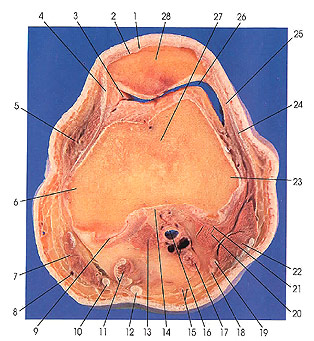
This section passes through the patella (28) and the medial (6) and lateral (23) epicondyles of the femur (27). The intercondylar fossa (14) and the articular cavity (26, 9) are seen.
At this level, several tendons and a retinaculum are found. These include the tendon of the vastus lateralis muscle (25), iliotibial tract (24), tendon of semimembranosus muscle (11), tendon of semitendinosus muscle (12), tendon of gracilis muscle (10), the transverse medial patellar retinaculum (4), and the tendon of the quadriceps muscle (2).
The lateral sural cutaneous nerve (18) (seen for the first time) and the posterior femoral cutaneous nerve (15) are identified.
The lateral sural cutaneous nerve is extremely variable. It leaves the common peroneal in the popliteal space and supplies the skin of the lower lateral part of the calf. One branch (peroneal anastomotic branch) may join with a branch of the medial sural cutaneous nerve to form the sural nerve. This nerve is also variable but usually descends along the lateral edge of the calcaneal tendon and sends lateral calcaneal branches to the lateral side of the heel, as well as a lateral dorsal cutaneous nerve to the lateral side of the foot.
The posterior femoral cutaneous nerve arises from the first, second, and third sacral nerves. It leaves the pelvis at the posterior surface of the sciatic nerve. It continues distally in the thigh, breaking up into terminal branches. Perineal branches go to the skin of the medial part of the thigh and the adjacent part of the scrotum or labium majus. One of these branches, the long pudendal nerve, runs medially dorsad to the ischial tuberosity. Inferior cluneal branches go to the skin of the lower medial part of the buttock. Femoral branches supply the skin of the back of the thigh and calf.
The alar plica (3) or alar folds are prominent crescentic folds of synovial membrane, extending dorsalward on each side of the patella from the base of the infrapatellar synovial fold. Their free margins are concave and thin.
Next Page | Previous Page | Section Top | Title Page
Please send us comments by filling out our Comment Form.
All contents copyright © 1995-2024 the Author(s) and Michael P. D'Alessandro, M.D. All rights reserved.
"Anatomy Atlases", the Anatomy Atlases logo, and "A digital library of anatomy information" are all Trademarks of Michael P. D'Alessandro, M.D.
Anatomy Atlases is funded in whole by Michael P. D'Alessandro, M.D. Advertising is not accepted.
Your personal information remains confidential and is not sold, leased, or given to any third party be they reliable or not.
The information contained in Anatomy Atlases is not a substitute for the medical care and advice of your physician. There may be variations in treatment that your physician may recommend based on individual facts and circumstances.
URL: http://www.anatomyatlases.org/