
Atlas of Human Anatomy in Cross Section: Section 7. Lower Limb
Ronald A. Bergman, Ph.D., Adel K. Afifi, M.D., Jean J. Jew, M.D., and Paul
C. Reimann, B.S.
Peer Review Status: Externally Peer Reviewed
|
Upper Left Quadrant |
Lower Left Quadrant |
Lower Right Quadrant |
Upper Right Quadrant |
|
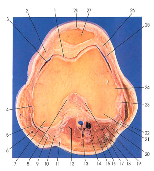
1. Articular cartilage |
4. Transverse collateral ligament |
13. Popliteal v. and a. |
25. Articular cavity |
This section passes through the lower third of the patella (27). It will not be seen in the next section. The section cuts several important ligaments and a retinaculum, including the lateral patellar (26), anterior cruciate (17), transverse collateral (4), and medial patellar (2).
Note that the sartorius muscle (5) has become a thin band as it crosses over and behind the medial condyle of the femur (7). All other "thigh" muscles have become tendinous--quadriceps femoris (28), biceps femoris (22), semitendinosus (11), semimembranosus (10), and gracilis (9).
The lateral patellar retinaculum (26) is derived from the tendon of vastus lateralis. It is attached to the lateral margin of the patella (nearer to the anterior than the posterior surface) as far as the junction with the patellar ligament, and passing along its sides to the tibia it inserts onto the oblique ridge. It extends as far as the fibular collateral ligament. The fibers of the lateral patellar retinaculum blend with the iliotibial tract of the fascia lata. The patellar retinacula (lateral and medial [21) become inseparably fused with the fibrous membrane of the articular capsule.
The fibular collateral ligament (23) is a strong round cord, about 5 cm long, attached proximally to a tubercle on the lateral epicondyle of the femur, just proximal and posterior to a groove from which the popliteus muscle arises. Distally, it is fixed to the middle of the lateral surface of the head of the fibula, about I cm or more anterior to the apex. Superficial to it lies the tendon of the biceps muscle (22), which bifurcates to surround the distal extremity of the ligament; deep to it are the popliteus tendon in its sheath, the inferior lateral (genicular) vessels of the knee, and a twig of the common peroneal nerve.
Next Page | Previous Page | Section Top | Title Page
Please send us comments by filling out our Comment Form.
All contents copyright © 1995-2024 the Author(s) and Michael P. D'Alessandro, M.D. All rights reserved.
"Anatomy Atlases", the Anatomy Atlases logo, and "A digital library of anatomy information" are all Trademarks of Michael P. D'Alessandro, M.D.
Anatomy Atlases is funded in whole by Michael P. D'Alessandro, M.D. Advertising is not accepted.
Your personal information remains confidential and is not sold, leased, or given to any third party be they reliable or not.
The information contained in Anatomy Atlases is not a substitute for the medical care and advice of your physician. There may be variations in treatment that your physician may recommend based on individual facts and circumstances.
URL: http://www.anatomyatlases.org/