
Atlas of Human Anatomy in Cross Section: Section 7. Lower Limb
Ronald A. Bergman, Ph.D., Adel K. Afifi, M.D., Jean J. Jew, M.D., and Paul
C. Reimann, B.S.
Peer Review Status: Externally Peer Reviewed
|
Upper Left Quadrant |
Lower Left Quadrant |
Lower Right Quadrant |
Upper Right Quadrant |
|
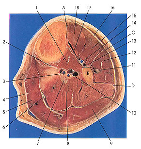
1. Tibialis posterior m. |
3. Posterior tibial vessels |
8. Soleus m. |
11. Superficial peroneal nerve (musculocutaneous nerve of leg) |
This section is three below the preceding one (3 cm).
The anterior musculature of the leg is composed of four muscles: tibialis anterior (18), extensor digitorum longus and extensor hallucis longus (15), and peroneus tertius (arises below this section). The lateral musculature consists of two muscles: peroneus longus and brevis (12) muscles. The posterior musculature consists of six muscles in two groups, superficial and deep. The superficial group includes gastrocnemius (5, 9), soleus (8), and plantaris (tendon, 6 and Plate 7.17, 12) muscles. The deep group of posterior musculature includes popliteus (see Plate 7.18, 2), flexor digitorum longus (see Plate 7.20, 2), and the tibialis posterior (1) muscles.
The three compartments, anterior, lateral, and posterior, can be seen on this plate. The anterior compartment of the leg is defined by a line beginning at A, running along the interosseous membrane to B, and extending onward to C. The lateral or peroneal compartment of the leg is defined by a continuous line joining C, B, and D. The posterior compartment is posterior and medial to a line joining points A, B, and D.
The anterior musculature consists of the following. Tibialis anterior (18) arises from the lateral surface of the tibia and adjacent interosseous membrane (17) in the proximal half of the leg. Its tendon passes over the front of the lower end of the tibia to insert on the medial surface of the first cuneiform and on the first metatarsal of the foot. Extensor digitorum longus (15) arises from the lateral condyle of the tibia, from the anterior surface of the fibula and adjacent interosseous membrane (17), and from the fascia of the leg near the tibial origin and gives rise to a tendon that passes over the front of the distal end of the tibia and sends tendons to the terminal phalanges of the four lateral toes. Extensor hallucis longus (15) is a narrow muscle that arises from the middle two-fourths of the anterior surface of the fibula near the interosseous crest and from the distal half of the interosseous membrane (17). The muscle gives rise to a tendon that extends over the ankle and inserts onto the base of the distal phalanx of the great toe. Peroneus tertius arises from the distal third of the anterior surface of the fibula, neighboring interosseous membrane, and anterior intermuscular septum. Its tendon passes laterally through the same osteofibrous canal in the same synovial sheath as extensor digitorum longus and terminates on the fifth metatarsal.
Next Page | Previous Page | Section Top | Title Page
Please send us comments by filling out our Comment Form.
All contents copyright © 1995-2024 the Author(s) and Michael P. D'Alessandro, M.D. All rights reserved.
"Anatomy Atlases", the Anatomy Atlases logo, and "A digital library of anatomy information" are all Trademarks of Michael P. D'Alessandro, M.D.
Anatomy Atlases is funded in whole by Michael P. D'Alessandro, M.D. Advertising is not accepted.
Your personal information remains confidential and is not sold, leased, or given to any third party be they reliable or not.
The information contained in Anatomy Atlases is not a substitute for the medical care and advice of your physician. There may be variations in treatment that your physician may recommend based on individual facts and circumstances.
URL: http://www.anatomyatlases.org/