
Atlas of Human Anatomy in Cross Section: Section 7. Lower Limb
Ronald A. Bergman, Ph.D., Adel K. Afifi, M.D., Jean J. Jew, M.D., and Paul
C. Reimann, B.S.
Peer Review Status: Externally Peer Reviewed
|
Upper Left Quadrant |
Lower Left Quadrant |
Lower Right Quadrant |
Upper Right Quadrant |
|
1. Tibialis anterior m. |
6. Tibial nerve |
9. Gastrocnemius m. aponeurosis (tendon m. gastrocnemius) |
13. Peroneus brevis |
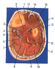
This section is three below the preceding one (3 cm).
No significant changes in structure have taken place, although the bulk of the belly of the soleus (7, 10) is becoming smaller as the sections proceed distally. The gastrocnemius muscle aponeurosis (8, 9) is well defined and thick. Note the tendon of plantaris (7) on the medial border of the aponeurosis of gastrocnemius (8).
The anterior tibial artery is the smaller of the two branches into which the popliteal artery divides at the distal border of the popliteus muscle. It courses between the tibia and fibula above the proximal part of the interosseous membrane, enters the anterior compartment, and runs distally in the anterior and lateral aspect of the leg, between the anterior muscles, as far as the anterior side of the ankle joint. Distal to this point the artery is renamed and is known as the dorsalis pedis artery. The artery is accompanied by a vena comitans. It is also accompanied, in the distal third of its course, by the deep peroneal nerve. This nerve, which winds around the head of the fibula and pierces the extensor digitorum longus, first joins the artery on its lateral side at about the proximal third of the leg. In the middle third it is anterior to the artery, and in the distal third it is again on the lateral side. The branches of the anterior tibial artery are the posterior tibial recurrent, anterior tibial recurrent, medial anterior malleolar, lateral anterior malleolar, and ten to fifteen muscular branches to the extensor of the toes and the anterior tibial muscle.
The posterior tibial artery is the larger of the two branches of the popliteal artery. It runs distally on the flexor aspect of the leg between the superficial and deep muscles and deep within the posterior compartment. Continuing obliquely downward to the tibial side of the leg, it reaches the ankle joint and passes deep to the flexor retinaculum between the flexor digitorum longus and flexor hallucis longus tendons, to the deep side of the origin of the abductor hallucis muscle, where it terminates by dividing into medial and lateral plantar arteries.
The branches of the posterior tibial artery are the fibular circumflex branch, peroneal, tibial nutrient, communicating posterior medial malleolar, and the medial calcaneal. It supplies muscular branches to those it passes, as well as to the skin of the lower medial region of the leg.
Next Page | Previous Page | Section Top | Title Page
Please send us comments by filling out our Comment Form.
All contents copyright © 1995-2024 the Author(s) and Michael P. D'Alessandro, M.D. All rights reserved.
"Anatomy Atlases", the Anatomy Atlases logo, and "A digital library of anatomy information" are all Trademarks of Michael P. D'Alessandro, M.D.
Anatomy Atlases is funded in whole by Michael P. D'Alessandro, M.D. Advertising is not accepted.
Your personal information remains confidential and is not sold, leased, or given to any third party be they reliable or not.
The information contained in Anatomy Atlases is not a substitute for the medical care and advice of your physician. There may be variations in treatment that your physician may recommend based on individual facts and circumstances.
URL: http://www.anatomyatlases.org/