
Atlas of Human Anatomy in Cross Section: Section 7. Lower Limb
Ronald A. Bergman, Ph.D., Adel K. Afifi, M.D., Jean J. Jew, M.D., and Paul
C. Reimann, B.S.
Peer Review Status: Externally Peer Reviewed
|
Upper Left Quadrant |
Lower Left Quadrant |
Lower Right Quadrant |
Upper Right Quadrant |
|
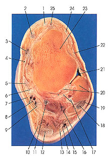
1. Tendon m. extensor hallucis longus |
5. Tendon m. tibialis posterior |
13. Sural nerve, lateral calcaneal br. |
21. Articular cavity and anterior talofibular ligament |
This section passes through the talus (23) just below the medial malleolus of the tibia. The lateral malleolus of the fibula (20) and its articulation with the talus are seen. The anterior (21) and posterior (20) talofibular ligaments are recognized. The dorsalis pedis artery (25) is seen for the first time in this section; it is the continuation of the anterior tibial artery. It extends from the ankle joint to the proximal end of the first interosseous space, where it divides into the first metatarsal and deep plantar arteries, which join the lateral plantar artery to complete the plantar arch. It lies along the lateral side of extensor hallucis longus muscle, and it is accompanied by a vena comitans and the deep peroneal nerve.
The peroneal artery has divided, and its posterior lateral malleolar and lateral calcaneal branches (16) are identified.
The lateral malleolus of the fibula is slightly flattened (side to side), and extends from the body (or shaft) of the fibula as an inverted pyramid. It is longer and more prominent than its medial counterpart (medial tibial malleolus). Its lateral surface is convex and subcutaneous. The medial surface has an anteroproximal malleolar articular surface, triangular in outline, convex, and articulating with the lateral side of the talus. The apex of the malleolus provides an attachment site for the calcaneofibular ligament. The anterior border of the malleolus is the attachment site for the anterior talofibular, anterior tibiofibular, and calcaneofibular ligaments. The posterior border is grooved in relation to the peroneal tendons (18, 19).
The talus (23) is the second largest of the bones of the tarsus. Superiorly it supports the tibia, inferiorly it rests upon the calcaneus, medially and laterally it articulates with the malleoli, and anteriorly it articulates with the navicular. The talus provides no attachment sites for muscle.
Next Page | Previous Page | Section Top | Title Page
Please send us comments by filling out our Comment Form.
All contents copyright © 1995-2024 the Author(s) and Michael P. D'Alessandro, M.D. All rights reserved.
"Anatomy Atlases", the Anatomy Atlases logo, and "A digital library of anatomy information" are all Trademarks of Michael P. D'Alessandro, M.D.
Anatomy Atlases is funded in whole by Michael P. D'Alessandro, M.D. Advertising is not accepted.
Your personal information remains confidential and is not sold, leased, or given to any third party be they reliable or not.
The information contained in Anatomy Atlases is not a substitute for the medical care and advice of your physician. There may be variations in treatment that your physician may recommend based on individual facts and circumstances.
URL: http://www.anatomyatlases.org/