
Atlas of Human Anatomy in Cross Section: Section 7. Lower Limb
Ronald A. Bergman, Ph.D., Adel K. Afifi, M.D., Jean J. Jew, M.D., and Paul
C. Reimann, B.S.
Peer Review Status: Externally Peer Reviewed
|
Upper Left Quadrant |
Lower Left Quadrant |
Lower Right Quadrant |
Upper Right Quadrant |
|
1. Tendon m. extensor hallucis longus |
8. Calcaneus |
16. Small saphenous v. |
23. Extensor digitorum brevis m. |
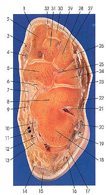
This section passes through the calcaneus (8, 18); the cuboid (24); navicular (5); the medial (first) (2), intermediate (second) (31), and lateral (third) (28) cuneiforms; and the second metatarsal (30). It cuts the cuneocuboid (25), plantar cuboideonavicular (25), interosseous talocalcaneal (21), plantar calcaneonavicular ("spring") (7), and medial (deltoid) (6) ligaments.
The plantar calcaneonavicular ligament (7) is a thick, dense plate of fibroelastic tissue. It extends from the sustentaculum tall and the distal surface of the calcaneus to the whole width of the inferior surface of the navicular bone. Medially, it blends with the anterior part of the medial ligament of the ankle joint and, laterally, with the plantar margin of the calcaneonavicular part of the bifurcate ligament. It is thickest at its medial margin (4 to 5 mm). In contact with the inferior surface of the ligament is the tendon of tibialis posterior muscle, supporting the head of the talus and thereby augmenting a function of the ligament. Because it is fibroelastic, the plantar calcaneonavicular ligament is also known as the "spring" ligament.
The plantar cuboideonavicular ligament (25) is a well-defined band of connective tissue that extends laterally from the plantar surface of the navicular to the depression on the medial surface of the cuboid, and slightly onto its plantar surface.
Next Page | Previous Page | Section Top | Title Page
Please send us comments by filling out our Comment Form.
All contents copyright © 1995-2025 the Author(s) and Michael P. D'Alessandro, M.D. All rights reserved.
"Anatomy Atlases", the Anatomy Atlases logo, and "A digital library of anatomy information" are all Trademarks of Michael P. D'Alessandro, M.D.
Anatomy Atlases is funded in whole by Michael P. D'Alessandro, M.D. Advertising is not accepted.
Your personal information remains confidential and is not sold, leased, or given to any third party be they reliable or not.
The information contained in Anatomy Atlases is not a substitute for the medical care and advice of your physician. There may be variations in treatment that your physician may recommend based on individual facts and circumstances.
URL: http://www.anatomyatlases.org/