
Atlas of Human Anatomy in Cross Section: Section 7. Lower Limb
Ronald A. Bergman, Ph.D., Adel K. Afifi, M.D., Jean J. Jew, M.D., and Paul
C. Reimann, B.S.
Peer Review Status: Externally Peer Reviewed
|
Upper Left Quadrant |
Lower Left Quadrant |
Lower Right Quadrant |
Upper Right Quadrant |
|
1. Tendon m. extensor digitorum longus |
9. Tendon m. extensor digitorum longus |
15. Calcaneal tendon (Achilles) |
23. Abductor hallucis m. |
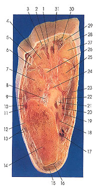
This is the superior (proximal) surface of the next section, looking distally. This section passes through the calcaneus (14); cuboid (7); the lateral (26) and medial (25) cuneiforms; and the first (27), second (31), third (4), and fourth (6) metatarsals. The dorsal metatarsal (2, 30), deep plantar (24), lateral and medial plantar (17, 20), and lateral malleolar (10) arteries are identified. The inferior peroneal retinaculum. (13) and the retrocalcaneal bursa (16) are seen for the first time.
The dorsal interosseous (30) muscles also make their first appearance. The dorsal interosseous muscles are larger than their plantar counterparts, are four in number, and fill the intermetatarsal spaces. The first two are inserted onto each side of the base of the proximal phalanx of the second toe; the third and fourth are inserted onto the lateral sides of the bases of the proximal phalanges of the third and fourth toes. In the foot, the axis about which the interosseous muscles are arranged passes through the second toe. The dorsal interossei abduct the toes from this axis. In the hand, however, the axis is the middle finger.
The inferior peroneal retinaculum 13) overlies the tendons of peroneus longus (13) and brevis (13) on the lateral surface of the calcaneus and is attached to this bone on each side of the tendons. Between the tendons it sends a septum to the bone. It is connected with the superficial layer of the inferior extensor retinaculum.
Next Page | Previous Page | Section Top | Title Page
Please send us comments by filling out our Comment Form.
All contents copyright © 1995-2024 the Author(s) and Michael P. D'Alessandro, M.D. All rights reserved.
"Anatomy Atlases", the Anatomy Atlases logo, and "A digital library of anatomy information" are all Trademarks of Michael P. D'Alessandro, M.D.
Anatomy Atlases is funded in whole by Michael P. D'Alessandro, M.D. Advertising is not accepted.
Your personal information remains confidential and is not sold, leased, or given to any third party be they reliable or not.
The information contained in Anatomy Atlases is not a substitute for the medical care and advice of your physician. There may be variations in treatment that your physician may recommend based on individual facts and circumstances.
URL: http://www.anatomyatlases.org/