
Lung
terminal bronchiole
Ronald A. Bergman, Ph.D., Adel K. Afifi, M.D., Paul M. Heidger,
Jr., Ph.D.
Peer Review Status: Externally Peer Reviewed
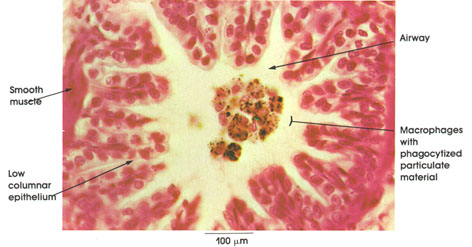
Cat, 10% formalin, H. & E., 324 x.
This illustration shows a cross section of a terminal bronchiole with phagocytized material (black) in macrophages within the lumen of the bronchiole (airway). The pleating of the epithelial lining denotes a constricted bronchiole. Note the low columnar epithelial lining of the wall of the bronchiole and the smooth muscle bundle adjacent to the lining epithelium. (See also Plate 226.)
Next Page | Previous Page | Section Top | Title Page
Please send us comments by filling out our Comment Form.
All contents copyright © 1995-2024 the Author(s) and Michael P. D'Alessandro, M.D. All rights reserved.
"Anatomy Atlases", the Anatomy Atlases logo, and "A digital library of anatomy information" are all Trademarks of Michael P. D'Alessandro, M.D.
Anatomy Atlases is funded in whole by Michael P. D'Alessandro, M.D. Advertising is not accepted.
Your personal information remains confidential and is not sold, leased, or given to any third party be they reliable or not.
The information contained in Anatomy Atlases is not a substitute for the medical care and advice of your physician. There may be variations in treatment that your physician may recommend based on individual facts and circumstances.
URL: http://www.anatomyatlases.org/