
Ronald A. Bergman, Ph.D., Adel K. Afifi, M.D., Paul M. Heidger,
Jr., Ph.D.
Peer Review Status: Externally Peer Reviewed
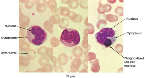
Human, air-dried blood smear, Wright's stain, 4416 x.
Monocytes are the largest cells found in normal blood. The nucleus is centrally or peripherally located, indented, and ovoid or horseshoe-shaped; the nuclear chromatin is not as dense as that of lymphocytes. Cytoplasm is abundant and contains azurophilic granules, which are usually smaller than those seen in lymphocytes. Monocytes are voracious phagocytes. The monocyte seen on the extreme right shows pseudopodia extending from the cell body and contains a phagocytized red cell nucleus
Note the comparative size of erythrocytes and monocytes.
Next Page | Previous Page | Section Top | Title Page
Please send us comments by filling out our Comment Form.
All contents copyright © 1995-2024 the Author(s) and Michael P. D'Alessandro, M.D. All rights reserved.
"Anatomy Atlases", the Anatomy Atlases logo, and "A digital library of anatomy information" are all Trademarks of Michael P. D'Alessandro, M.D.
Anatomy Atlases is funded in whole by Michael P. D'Alessandro, M.D. Advertising is not accepted.
Your personal information remains confidential and is not sold, leased, or given to any third party be they reliable or not.
The information contained in Anatomy Atlases is not a substitute for the medical care and advice of your physician. There may be variations in treatment that your physician may recommend based on individual facts and circumstances.
URL: http://www.anatomyatlases.org/