
Ronald A. Bergman, Ph.D., Adel K. Afifi, M.D., Paul M. Heidger,
Jr., Ph.D.
Peer Review Status: Externally Peer Reviewed
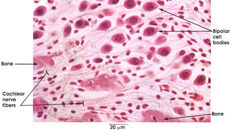
Guinea pig, Müller's fluid, H. & E., 612 x.
Bone: Modiolus or central conical pillar of spongy bone of the osseous cochlea.
Cochlear nerve fibers: Central processes of the bipolar ganglion cells.
Bipolar cell bodies: Spiral ganglion. Peripheral processes of sensory hair cells located in the organ of Corti.* Central processes from the spiral ganglion cells form the cochlear nerve (auditory part of the eighth cranial nerve).
See Plate 311.
*Corti was a nineteenth-century Italian anatomist.
Next Page | Previous Page | Section Top | Title Page
Please send us comments by filling out our Comment Form.
All contents copyright © 1995-2024 the Author(s) and Michael P. D'Alessandro, M.D. All rights reserved.
"Anatomy Atlases", the Anatomy Atlases logo, and "A digital library of anatomy information" are all Trademarks of Michael P. D'Alessandro, M.D.
Anatomy Atlases is funded in whole by Michael P. D'Alessandro, M.D. Advertising is not accepted.
Your personal information remains confidential and is not sold, leased, or given to any third party be they reliable or not.
The information contained in Anatomy Atlases is not a substitute for the medical care and advice of your physician. There may be variations in treatment that your physician may recommend based on individual facts and circumstances.
URL: http://www.anatomyatlases.org/