
Ronald A. Bergman, Ph.D., Adel K. Afifi, M.D., Paul M. Heidger,
Jr., Ph.D.
Peer Review Status: Externally Peer Reviewed

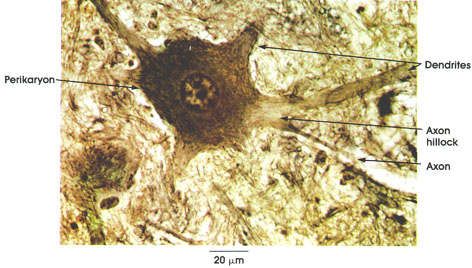
Rhesus monkey, 10% formalin, Glees' method, 612 x.
Perikaryon: It is multipolar (i.e., possesses a single axon and several dendrites) and has a central prominent nucleus. Cytoplasm is rich in Nissl* bodies except at the axon hillock (lighter area of cell body from which axon arises). Nissl bodies can be seen in Plates 1 and 90.
Dendrites: Stout tapering processes similar in structure to the perikaryon.
Axon: This arises from the axon hillock. It is a slender process of uniform diameter and great length. The myelin sheath is not seen around the axon in this preparation because it is not preserved by the fixation method used.
*Nissl was a nineteenth-century German neurologist.
Next Page | Previous Page | Section Top | Title Page
Please send us comments by filling out our Comment Form.
All contents copyright © 1995-2024 the Author(s) and Michael P. D'Alessandro, M.D. All rights reserved.
"Anatomy Atlases", the Anatomy Atlases logo, and "A digital library of anatomy information" are all Trademarks of Michael P. D'Alessandro, M.D.
Anatomy Atlases is funded in whole by Michael P. D'Alessandro, M.D. Advertising is not accepted.
Your personal information remains confidential and is not sold, leased, or given to any third party be they reliable or not.
The information contained in Anatomy Atlases is not a substitute for the medical care and advice of your physician. There may be variations in treatment that your physician may recommend based on individual facts and circumstances.
URL: http://www.anatomyatlases.org/