
Ronald A. Bergman, Ph.D., Adel K. Afifi, M.D., Paul M. Heidger,
Jr., Ph.D.
Peer Review Status: Externally Peer Reviewed
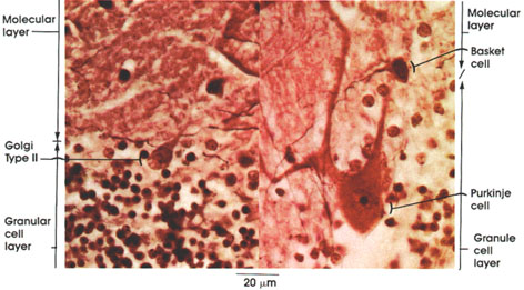
Mouse,
20% formalin,
Bielschowsky's method, 612 x.
Molecular layer: Most superficial layer of the cerebellar cortex. It is sparsely cellular and largely synaptic.
Basket cell: A large variety of stellate cells deep in the molecular layer in the vicinity of Purkinje cells. Axons run transversely in the molecular layer and send collaterals that arborize around the perikarya of Purkinje cells like a basket.
Purkinje cell: Single row of large, flask-shaped cells. Form a distinct layer bordering the molecular and granule cell layers. Dendrites arborize richly in the molecular layer. These cells are named after Johannes Purkinje, a Bohemian physiologist, who described them in 1837.
Granule cell layer: Closely packed with chromatic nuclei of small granule cell neurons. Major input to the cerebellum projects into this layer.
Golgi Type II: This type of cerebellar neuron is found in the upper part of the granule cell layer close to the Purkinje cell layer. Larger than the granule cell neuron. Dendrites arborize extensively in the molecular layer. Axons establish synapses with dendrites of granule cells in the glomeruli of the granule cell layer. It is estimated that there is one Golgi Type II cell for every 10 Purkinje cells.
Next Page | Previous Page | Section Top | Title Page
Please send us comments by filling out our Comment Form.
All contents copyright © 1995-2024 the Author(s) and Michael P. D'Alessandro, M.D. All rights reserved.
"Anatomy Atlases", the Anatomy Atlases logo, and "A digital library of anatomy information" are all Trademarks of Michael P. D'Alessandro, M.D.
Anatomy Atlases is funded in whole by Michael P. D'Alessandro, M.D. Advertising is not accepted.
Your personal information remains confidential and is not sold, leased, or given to any third party be they reliable or not.
The information contained in Anatomy Atlases is not a substitute for the medical care and advice of your physician. There may be variations in treatment that your physician may recommend based on individual facts and circumstances.
URL: http://www.anatomyatlases.org/