
Ronald A. Bergman, Ph.D., Adel K. Afifi, M.D., Paul M. Heidger,
Jr., Ph.D.
Peer Review Status: Externally Peer Reviewed
Human, Helly's fluid, toluidine blue and erythrosin stains, 162 x.
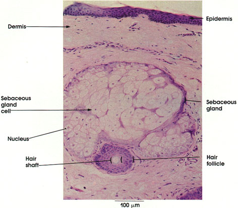
Epidermis: Stratified squamous cornified epithelium of the skin.
Dermis: Connective tissue layer beneath the epidermis. Its thickness varies in different parts of the body. It is rich in collagenous and elastic fibers. The part of the dermis underlying the epithelium is called the papillary layer. The deeper part is the reticular layer, in which sebaceous glands are found. In addition, hair follicles, sweat glands, and Pacinian corpuscles occur in this layer. In the face, the striated muscles of facial expression terminate in the dermis.
Sebaceous gland: Holocrine variety of gland in which the entire cell is lost along with the secretory products. Intimately associated with hair follicles into which they drain. Composed of a group of saclike alveoli ensheathed by a thin layer of connective tissue. The alveoli are composed of stratified cuboidal or polyhedral epithelia[ cells that fill the sac. The secretion of the sebaceous gland is an oily substance (sebum) that lubricates the epidermis and hair.
Sebaceous gland cell: Note the peripheral, small cuboidal cells and the more central, larger polyhedral or spheroidal cells. Oily droplets increase with an increase in size of the cells. See Plate 83.
Nucleus: Nuclei of peripheral cells are rounded. Nuclei of centrally located cells are either shrunken or absent. This nuclear change is part of the degenerative process by which the entire cell is lost, along with its secretion product.
Hair follicle: Surrounds the hair shaft and is composed of inner epidermal epithelial elements and outer dermal connective tissue elements.
Hair shaft: Located within the follicle. The free end of the hair projects from the surface of the skin.
Next Page | Previous Page | Section Top | Title Page
Please send us comments by filling out our Comment Form.
All contents copyright © 1995-2024 the Author(s) and Michael P. D'Alessandro, M.D. All rights reserved.
"Anatomy Atlases", the Anatomy Atlases logo, and "A digital library of anatomy information" are all Trademarks of Michael P. D'Alessandro, M.D.
Anatomy Atlases is funded in whole by Michael P. D'Alessandro, M.D. Advertising is not accepted.
Your personal information remains confidential and is not sold, leased, or given to any third party be they reliable or not.
The information contained in Anatomy Atlases is not a substitute for the medical care and advice of your physician. There may be variations in treatment that your physician may recommend based on individual facts and circumstances.
URL: http://www.anatomyatlases.org/