
Ronald A. Bergman, Ph.D., Adel K. Afifi, M.D., Paul M. Heidger,
Jr., Ph.D.
Peer Review Status: Externally Peer Reviewed
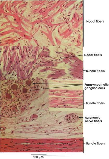
The atrioventricular (AV) node and bundle are composed of cardiac muscle fibers specialized for impulse conduction. The AV node (node of Tawara*) is found in the subendocardium of the right atrium, close to the termination of the coronary sinus. Note the irregularly arranged branching fibers (nodal fibers) that form the AV node. Note also how the AV nodal fibers become continuous with the small unbranched fibers of the AV bundle (bundle of His). The latter originate in the AV node, continue into the interventricular septum, and divide into two trunks composed of Purkinje fibers, which pass to the right and left ventricular wall, where they become continuous with ordinary cardiac muscle fibers. Stimuli for cardiac contraction are initiated in the sinoatrial (SA) node, reach the AV node, and spread to the myocardium via the AV bundle. Injury to the bundle results in dissociation of atrial and ventricular rhythms.
Note the parasympathetic ganglion cells and the autonomic nerve fibers in the wall of the heart. The parasympathetic ganglia receive vagal fibers, which slow the heart rate while the sympathetic postganglionic fibers carry impulses that increase the heart rate. The autonomic fibers include sympathetic postganglionic and parasympathetic pre- and postganglionic fibers.
*Tawara, 1873-1952, was a Japanese physician.
Next Page | Previous Page | Section Top | Title Page
Please send us comments by filling out our Comment Form.
All contents copyright © 1995-2024 the Author(s) and Michael P. D'Alessandro, M.D. All rights reserved.
"Anatomy Atlases", the Anatomy Atlases logo, and "A digital library of anatomy information" are all Trademarks of Michael P. D'Alessandro, M.D.
Anatomy Atlases is funded in whole by Michael P. D'Alessandro, M.D. Advertising is not accepted.
Your personal information remains confidential and is not sold, leased, or given to any third party be they reliable or not.
The information contained in Anatomy Atlases is not a substitute for the medical care and advice of your physician. There may be variations in treatment that your physician may recommend based on individual facts and circumstances.
URL: http://www.anatomyatlases.org/